中国组织工程研究 ›› 2020, Vol. 24 ›› Issue (9): 1464-1470.doi: 10.3969/j.issn.2095-4344.2516

• 骨与关节循证医学 evidence-based medicine of the bone and joint • 上一篇    下一篇

显微镜辅助与传统直视下颈椎前路减压治疗颈椎病的Meta分析

罗海涛,程祖珏,吕世刚,肖爵贤,何  伟,黄  凯,范阳华,祝新根   

  1. 南昌大学第二附属医院神经外科,江西省南昌市  330006
  • 收稿日期:2019-08-10 修回日期:2019-08-14 接受日期:2019-10-09 出版日期:2020-03-28 发布日期:2020-02-13
  • 通讯作者: 程祖珏,教授,主任医师,南昌大学第二附属医院神经外科,江西省南昌市 330000
  • 作者简介:罗海涛,男,1993年生,汉族,2019年南昌大学毕业,硕士,主要从事神经外科脊柱脊髓亚专业的研究。
  • 基金资助:
    江西省教育厅科学技术研究项目(60159);江西省卫生与计划生育管理委员会项目(20175224);卫生部医药卫生科技发展研究中心项目(W2014ZT268);江西省卫生与计划生育管理委员会项目(20165215)

Microscope-assisted versus traditional anterior cervical approach for cervical spondylopathy: a meta-analysis

Luo Haitao, Cheng Zujue, Lü Shigang, Xiao Juexian, He Wei, Huang Kai, Fan Yanghua, Zhu Xingen   

  1. Department of Neurosurgery, Second Affiliated Hospital of Nanchang University, Nanchang 330006, Jiangxi Province, China
  • Received:2019-08-10 Revised:2019-08-14 Accepted:2019-10-09 Online:2020-03-28 Published:2020-02-13
  • Contact: Cheng Zujue, Professor, Chief physician, Department of Neurosurgery, Second Affiliated Hospital of Nanchang University, Nanchang 330006, Jiangxi Province, China
  • About author:Luo Haitao, Master, Department of Neurosurgery, Second Affiliated Hospital of Nanchang University, Nanchang 330006, Jiangxi Province, China
  • Supported by:
    the Science and Technology Program of Education Department of Jiangxi Province, No. 60159; the Project of Health and Family Planning Commission of Jiangxi Province, No. 20175224; the Project of Development Center for Medical and Health Science and Technology of the Ministry of Health of China, No. W2014ZT268; the Project of Health and Family Planning Commission of Jiangxi Province, No. 20165215

摘要:

文题释义:
颈椎前路椎间盘切除减压:一度成为治疗颈椎退行性疾病的主流术式,然而在实际手术操作过程中,由于狭小的手术视野等原因,使得术区的解剖结构常不能被清晰分辨,进而加大了对受压脊髓和神经根减压的难度。
显微辅助下行颈椎前路减压:1975年Hankinson等就首次报道了显微镜辅助下前路椎间盘切除减压融合术,目前在欧美国家已成为神经外科与脊柱外科的标准术式之一,但在国内仍需进一步推广。

背景:虽然近年来国内不少脊柱外科医师开始使用显微镜行颈前路减压手术,但显微镜的使用仍未在国内推广开来。因此有必要对显微镜辅助颈椎前路精细化减压与传统直视下颈椎前路减压治疗颈椎病的临床疗效进行系统评价和分析,为颈椎病治疗的临床决策提供证据支持。

目的:系统评价显微镜辅助与传统直视下颈椎前路减压治疗颈椎病的临床疗效。

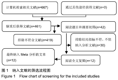
方法:检索Medline、Embase、PubMed、Web of Science、中国生物医学文献数据库(CBM)、万方、维普以及中国知网等数据库自建库以来至2019年7月有关显微镜辅助与传统直视下行颈椎前路减压治疗颈椎病疗效对比的文献,由2名研究人员独立进行文献筛选。并对2组手术时间、术中出血量、术前及术后日本骨科协会评分、神经功能改善率、术前及术后目测类比评分、并发症发生率等指标进行比较。采用国际Cochrane协作网推荐的改良Jadad量表、改良纽卡斯尔-渥太华量表及MINORS量表对纳入研究的文献进行质量评价,采用Stata 12.0软件进行对相关结局指标进行Meta分析。

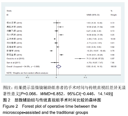
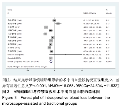
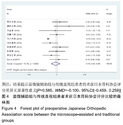
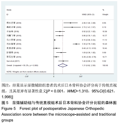
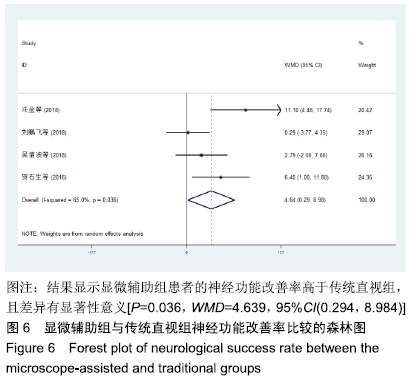
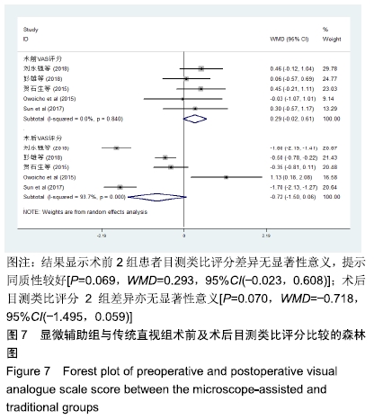
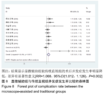
结果与结论:①12篇研究符合纳入标准,共包括892例患者,其中显微辅助组438例,传统直视组454例;②显微辅助组与传统直视组患者的术前日本骨科协会评分[WMD=-0.100,95%CI(-0.459,0.259),P=0.585]、手术时间[WMD=6.852,95%CI(-0.446,14.149),P=0.066]、术前及术后目测类比评分[WMD=0.293,95%CI(-0.023,0.608),P=0.069;WMD=-0.718,95%CI(-1.495,0.059),P=0.070]相比,差异均无显著性意义;③与传统直视组相比,显微辅助组能显著提高患者的术后日本骨科协会评分[WMD=1.310,95%CI(0.621,1.998),P < 0.001]和神经功能改善率[WMD=4.639,95%CI(0.294,8.984),P=0.036],并能显著减少术中出血量[WMD=-18.068,95%CI(-24.504,-11.632),P < 0.001],降低并发症发生率[RR=1.068,95%CI(1.012,1.126),P=0.002];④提示与传统直视下手术相比,显微镜辅助下行颈椎前路减压治疗颈椎病具有更好的临床疗效,但仍需要更多大样本、高质量的临床研究加以验证。

ORCID: 0000-0002-9190-1386(罗海涛)

中国组织工程研究杂志出版内容重点:人工关节;骨植入物;脊柱骨折;内固定;数字化骨科;组织工程


关键词: 显微镜辅助, 颈椎前路减压, 颈椎病, 传统直视, Meta分析

Abstract:

BACKGROUND: In China, although microscope-assisted anterior cervical decompression has been performed by many spine surgeons in recent years, the application of microscope still needs to be further promoted in China. Therefore, it is necessary to systematically evaluate and analyze the clinical efficacy of cervical spondylopathy by comparing patients under microscope-assisted anterior cervical approach to traditional anterior cervical approach. As a result, it can provide the evidence for clinical decision in these cervical spondylopathy patients.

OBJECTIVE: To systematically evaluate the effect between microscope-assisted anterior cervical approach and traditional anterior cervical approach in cervical spondylopathy patients.

METHODS: Databases such as Medline, Embase, PubMed, Web of science, CBM, Wangfang databases, VIP, and CNKI were searched from inception to July 2019 to find literature related to microscope-assisted anterior cervical approach and traditional anterior cervical approach in treatment of cervical spondylopathy. Two investigators evaluated the researches independently and compared the operative time, intraoperative blood loss, the preoperative and postoperative Japanese Orthopedic Association score, neurological success rate, the preoperative and the postoperative visual analogue scale score, and the complication rates between two groups. The quality of the included literature was evaluated by the modified Jadad scale, the modified Newcastle Ottawa score and the methodological index for non-randomized-studies. The correlative clinical outcome in inclusive research was analyzed systematically by using Stata 12.0 Software.

RESULTS AND CONCLUSION: (1) Twelve studies were accorded with the inclusion criteria, containing 892 cases (438 cases in the microscope-assisted group, while 454 cases in the traditional group). (2) There were no significant differences in the preoperative Japanese Orthopedic Association score (WMD=-0.100, 95%CI(-0.459, 0.259), P=0.585), operative time (WMD=6.852, 95%CI(-0.446, 14.149), P=0.066), the preoperative and postoperative visual analogue scale score (WMD=0.293, 95%CI(-0.023, 0.608), P=0.069; WMD=-0.718, 95%CI(-1.495, 0.059), P=0.070) between two groups. (3) Postoperative Japanese Orthopedic Association score (WMD=1.310, 95%CI(0.621, 1.998), P < 0.001), the rate of neurological success (WMD=4.639, 95%CI(0.294, 8.984), P=0.036) in the microscope-assisted group were higher than those in the traditional group. The intraoperative blood loss (WMD=-18.068, 95%CI(-24.504, -11.632), P < 0.001) and the rate of complication (RR=1.068, 95%CI(1.012, 1.126), P=0.002) in the microscope-assisted group were significantly lower than those in the traditional group. (4) Performing anterior cervical approach under microscope has a better clinical effect than using traditional anterior cervical approach in the treatment of cervical spondylopathy, but more high-quality clinical studies are needed to verify it. 

Key words: microscope-assisted, anterior approach, cervical spondylopathy, traditional direct view, meta-analysis

中图分类号: