|
[1] ORBACH SM, LESS RR, KOTHARI A, et al. In Vitro Intestinal and Liver Models for Toxicity Testing.ACS Biomater Sci Eng. 2017;3(9):1898-1910.
[2] TAŠKOVA K, FONTAINE JF, MROWKA R, et al. Evaluation of in vivo and in vitro models of toxicity by comparison of toxicogenomics data with the literature.Methods. 2017;132: 57-65.
[3] NIEMAN GF, SATALIN J, ANDREWS P, et al. Preemptive mechanical ventilation based on dynamic physiology in the alveolar microenvironment: Novel considerations of time-dependent properties of the respiratory system.J Trauma Acute Care.2018;85(6):1081-1091.
[4] LI W, YAN Z, REN J, et al. Manipulating cell fate: dynamic control of cell behaviors on functional platforms.Chem Soc Rev2018;47(23):8639-8684.
[5] YAMAGISHI M, HORI Y, UEMURA S, et al. Time evolution of microenvironment around cells regulated by the secretion activity and culture density of the cells//International Symposium on Micro-nanomechatronics & Human Science. IEEE,2018.
[6] MORONI L, BURDICK JA, HIGHLEY C, et al. Biofabrication strategies for 3D in vitro models and regenerative medicine. Nat Rev Mater.2018;3(5):21-37.
[7] RYAN AJ, BROUGHAM CM, GARCIARENA CD, et al. Towards 3D in vitro models for the study of cardiovascular tissues and disease.Drug Discov Today.2016; 21(9):1437-1445.
[8] FIGTREE GA, BUBB KJ, TANG O, et al. Vascularized Cardiac Spheroids as Novel 3D in vitro Models to Study Cardiac Fibrosis.Cells Tissues Organs.2017; 204(3-4):191-198.
[9] FERRARINI M, STEIMBERG N, BONIOTTI J, et al. 3D-Dynamic Culture Models of Multiple Myeloma.Methods Mol Biol.2017;1612:177-190.
[10] JIN L, XU Q, KUDDANNAYA S, et al. Fabrication and Characterization of Three Dimensional Core-Shell Structure Nanofibers Designed for 3D Dynamic Cell Culture.ACS Appl Mater InteR.2017;9(21):17718-17726.
[11] PENDERECKA K, IBBS M, KALUZNA A, et al. Implementation of a dynamic culture condition to the heterotypic 3D breast cancer model.J Biomed Mater Res B.2019; 8:1-12.
[12] JARVIS M, ARNOLD M, OTT J, et al. Microfluidic co-culture devices to assess penetration of nanoparticles into cancer cell mass.Bioeng Transl Med.2017;2(3):268-277.
[13] ZHAO Y, YAN X, LI B, et al. Three-dimensional co-culture microfluidic model and its application for research on cancer stem-like cells inducing migration of endothelial cells. Biotechnol Lett.2017;39(9):1425-432.
[14] LI R, ZHANG X, LV X, et al. Microvalve controlled multi-functional microfluidic chip for divisional cell co-culture. Anal Biochem.2017;539:48-53.
[15] MASSAI D, BOLESANI E, DIAZ DR, et al. Sensitivity of human pluripotent stem cells to insulin precipitation induced by peristaltic pump-based medium circulation: considerations on process development OPEN.Sci Rep-UK.2017;7(1):3950.
[16] YEH SL, LIN TR, PENG CC, et al. A microfluidic device to study effects of physical stimulation and steroid treatment on lung epithelial cell surfactant protein expression//International Conference on Solid-state Sensors.IEEE, 2017.
[17] LI J, WEI J, LIU Y, et al. A microfluidic design to provide a stable and uniform in vitro microenvironment for cell culture inspired by the redundancy characteristic of leaf areoles.Lab Chip. 2017;17(22):3921-3933.
[18] WANG X, ZHAO D, PHAN DTT, et al. A hydrostatic pressure- driven passive micropump enhanced with siphon-based autofill function.Lab Chip. 2018;18(15):2167-2177.
[19] WANG YN, FU LM. Micropumps and biomedical applications- A review. Microelectron Eng.2018;195:121-138.
[20] HOROBIN JT, SIMMONDS MJ, NANDAKUMAR D, et al. Speed Modulation of the HeartWare HVAD to Assess In Vitro Hemocompatibility of Pulsatile and Continuous Flow Regimes in a Rotary Blood Pump.Artif Organs.2018;42(9):879-890.
[21] JOOHYUNG L, ZACHARY E, HIMALI S, et al. A microfluidic cardiac flow profile generator for studying the effect of shear stress on the valvular endothelial cell.Lab Chip. 2018;18(19): 2946-2956.
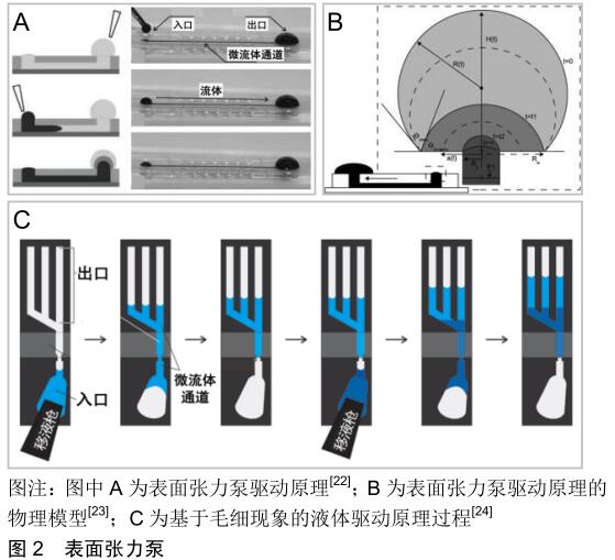
[22] MEYVANTSSON I, WARRICK JW, HAYES S, et al. Automated cell culture in high density tubeless microfluidic device arrays. Lab Chip.2008;8(5):717-724.
[23] BERTHIER E, BEEBE DJ. Flow rate analysis of a surface tension driven passive micropump.Lab Chip. 2007;7(11): 1475-1478.
[24] JUNCKER D, SCHMID H, DRECHSLER U, et al. Autonomous microfluidic capillary system.Anal Chem. 2002;74(24):6139-6144.
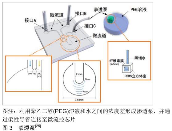
[25] PARK JY, YOO SJ, HWANG CM, et al. Simultaneous generation of chemical concentration and mechanical shear stress gradients using microfluidic osmotic flow comparable to interstitial flow.Lab Chip.2009;9(15):2194-2202.
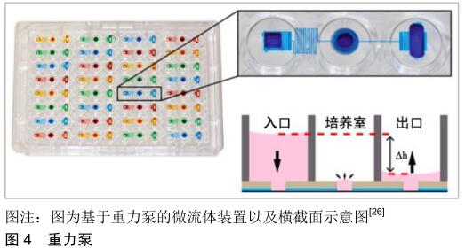
[26] CHEN SYC, HUNG PJ, LEE PJ. Microfluidic array for three-dimensional perfusion culture of human mammary epithelial cells.Biomed Microdevices.2011;13(4):753-758.
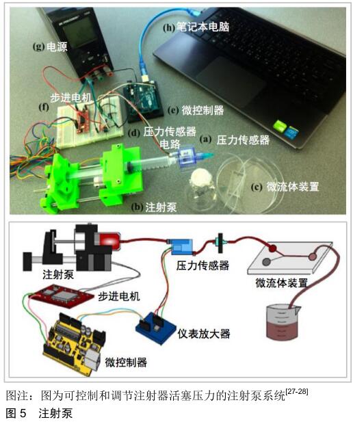
[27] CHEN MC, LAKE JR, HEYDE KC, et al. Three-dimensional Printing of Thermoplastic Materials to Create Automated Syringe Pumps with Feedback Control for Microfluidic Applications. Jove-J Vis Exp.2018;138:e57553.
[28] LAKE JR, HEYDE KC, RUDER WC. Low-cost feedback- controlled syringe pressure pumps for microfluidics applications. PLoS One.2017;12(4):e0175089.
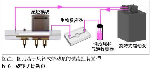
[29] SHAEGH SAM, FERRARI FD, ZHANG YS, et al. A microfluidic optical platform for real-time monitoring of pH and oxygen in microfluidic bioreactors and organ-on-chip devices. Biomicrofluidics.2016;10(4):044111.
[30] SKARDAL A, MURPHY SV, DEVARASETTY M, et al. Multi-tissue interactions in an integrated three-tissue organ-on-a-chip platform.Sci Rep-UK.2017;7(1):8837.
[31] SMITS JG. Piezoelectric micropump with three valves working peristaltically. Sensor Actuat A-Phys.1990;21(1-3):203-206.
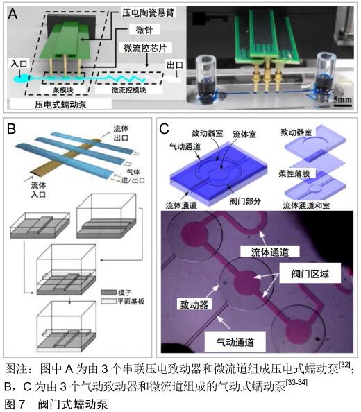
[32] MA T, SUN S, LI B, et al. Piezoelectric peristaltic micropump integrated on a microfluidic chip.Sensor Actuat A-Phys. 2019; 292:90-96.
[33] UNGER MA, CHOU HP, THORSEN T, et al. Monolithic Microfabricated Valves and Pumps by Multilayer Soft Lithography. Science.2000;288(5463):113-116.
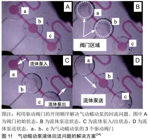
[34] JEONG OC, KONISHI S. Fabrication of a peristaltic micro pump with novel cascaded actuators.J Micromech Microeng. 2008;18(2):025022.
[35] PARK JY, KIM SK, WOO DH, et al. Differentiation of Neural Progenitor Cells in a Microfluidic Chip‐Generated Cytokine Gradient.Stem Cells.2009; 27(11):2646-2654.
[36] CHO BS, SCHUSTER TG, ZHU X, et al. Passively Driven Integrated Microfluidic System for Separation of Motile Sperm. Anal Chem.2003;75(7):1671-1675.
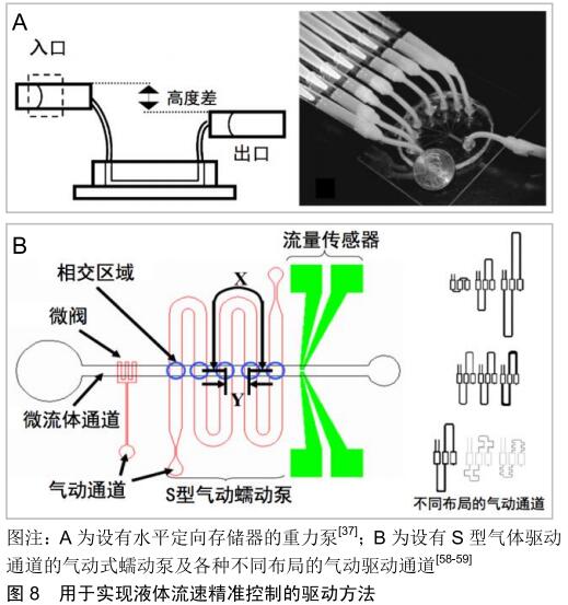
[37] ZHU XY, CHU LY, CHUEH BH, et al. Arrays of horizontally- oriented mini-reservoirs generate steady microfluidic flows for continuous perfusion cell culture and gradient generation. Analyst.2004;129(11):1026-1031.
[38] PARK JY, MORGAN M, SACHS AN, et al. Single cell trapping in larger microwells capable of supporting cell spreading and proliferation.Microfluid Nanofluid.2010;8(2):263-268.
[39] HUNG PJ, LEE PJ, SABOUNCHI P, et al. Continuous perfusion microfluidic cell culture array for high-throughput cell-based assays.Biotechnol Bioeng.2005;89(1):1-8.
[40] WANG J, HEO J, HUA SZ, et al. Spatially resolved shear distribution in microfluidic chip for studying force transduction mechanisms in cells.Lab Chip. 2009;10(2):235-239.
[41] VINAYAKUMAR KB, NADIGER G, SHETTY VR, et al. Packaged peristaltic micropump for controlled drug delivery application. Rev Sci Instrum.2017;88(1):015102.
[42] CHENG CH, YANG AS, LIN CJ, et al. Characteristic studies of a novel piezoelectric impedance micropump.Microsyst Technol.2017;23(6):1709-1717.
[43] POURMAND A, SHAEGH SAM, GHAVIFEKR HB, et al. Fabrication of Whole-Thermoplastic Normally Closed Microvalve, Micro Check Valve, and Micropump.Sensor Actuat B-Chem.2017;262:625-636.
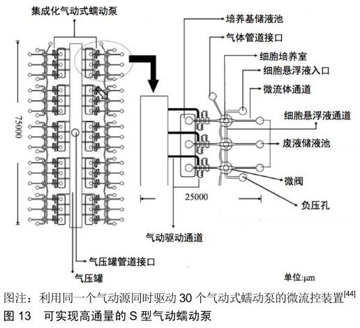
[44] WU MH, HUANG SB, CUI Z, et al.A high throughput perfusion-based microbioreactor platform integrated with pneumatic micropumps for three-dimensional cell culture. Biomed Microdevices.2008;10(2):309-319.
[45] AU AK, BHATTACHARJEE N, HOROWITZ LF, et al. 3D-printed microfluidic automation.Lab Chip.2015;15(8): 1934-1941.
[46] LEE YS, BHATTACHARJEE N, FOLCH A, et al. 3D-Printed Quake-Style Microvalves and Micropumps.Lab Chip. 2018; 18(8):1207-1214.
[47] ARORA S, LAM AJY, CHEUNG C, et al. Determination of critical shear stress for maturation of human pluripotent stem cell derived endothelial cells towards an arterial subtype. Biotechnol Bioeng.2018;116(5):1164-1175.
[48] WANG XL, ZHANG YY, FENG T, et al Fluid Shear Stress Promotes Autophagy in Hepatocellular Carcinoma Cells.Int J Biol Sci.2018;14(10):1277-1290.
[49] MEZA D, MUSMACKER B, STEADMAN E, et al. Endothelial Cell Biomechanical Responses are Dependent on Both Fluid Shear Stress and Tensile Strain. Cell Mol Bioeng. 2019;12(4): 311-325.
[50] YU L, MA X, SUN J, et al. Fluid shear stress induces osteoblast differentiation and arrests the cell cycle at the G0 phase via the ERK1/2 pathway.Mol Med Rep.2017;16(6):8699-8708.
[51] LANDWEHR GM ,KRISTOF AJ, RAHMAN SM, et al. Biophysical analysis of fluid shear stress induced cellular deformation in a microfluidic device.Biomicrofluidics. 2018; 12(5):054109.
[52] DELON LC, GUO ZB, OSZMIANA A, et al. A systematic investigation of the effect of the fluid shear stress on Caco-2 cells towards the optimization of epithelial organ-on-chip models. Biomaterials.2019;225:119521.
[53] BIROL S, FUCUCUOGLU R, CADIRCI S, et al. Investigation on the effects of variable shear stress on monocyte cell morphology.Micro Nano Lett.2017; 12(11):881-885.
[54] HUH D, MATTHEWS BD, MAMMOTO A, et al. Reconstituting Organ-Level Lung Functions on a Chip.Science. 2010;328 (5986):1662-1668.
[55] YAN Z, SU G, GAO W, et al. Fluid shear stress induces cell migration and invasion via activating autophagy in HepG2 cells.Cell Adhes Migr.2019;13(1):152-162.
[56] PRZYBYLA LM, VOLDMAN J. Attenuation of extrinsic signaling reveals the importance of matrix remodeling on maintenance of embryonic stem cell self-renewal.P Natl Acad Sci USA.2012;109(3):835-840.
[57] ESTRADA R, GIRIDHARAN GA, NGUYEN MD, et al. Endothelial Cell Culture Model for Replication of Physiological Profiles of Pressure, Flow, Stretch, and Shear Stress in Vitro. Anal Chem.2011;83(8):3170-3177.
[58] WANG CH, LEE GB. Pneumatically driven peristaltic micropumps utilizing serpentine-shape channels.J Micromech Microeng.2006;16(2):341-348.
[59] HUANG SB, WU MH, CUI Z, et al. A membrane-based serpentine-shape pneumatic micropump with pumping performance modulated by fluidic resistance.J Micromech Microeng.2008;18(4):045008.
[60] SOENKSEN LR, KASSIS T, NOH M, et al. Closed-loop feedback control for microfluidic systems through automated capacitive fluid height sensing.Lab Chip.2018;18(6):902-914.
[61] KANG YJ. Periodic and simultaneous quantification of blood viscosity and red blood cell aggregation using a microfluidic platform under in-vitro closed-loop circulation. Biomicrofluidics. 2018;12(2):024116.
[62] SUNG JH, KAM C, SHULER ML, et al. A microfluidic device for a pharmacokinetic–pharmacodynamic (PK-PD) model on a chip.Lab Chip.2010;10(4):446-455.
[63] ZHENG W, JIANG B, WANG D, et al. A microfluidic flow-stretch chip for investigating blood vessel biomechanics. Lab Chip. 2012;12(18):3441-3450.
[64] LANGILLE BL, ADAMSON SL. Relationship between blood flow direction and endothelial cell orientation at arterial branch sites in rabbits and mice.Circ Res.1981;48(4):481-488.
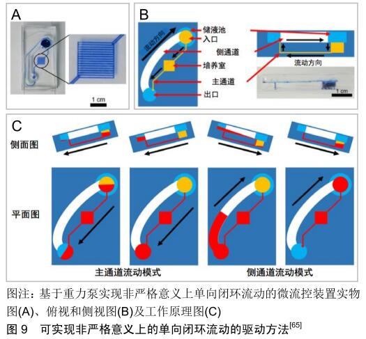
[65] LEE DW, CHOI N, SUNG JH. A microfluidic chip with gravity-induced unidirectional flow for perfusion cell culture. Biotechnol Progr.2019;35(1): UNSP e2701.
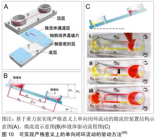
[66] WANG YI, SHULER ML. UniChip enables long-term recirculating unidirectional perfusion with gravity-driven flow for microphysiological systems.Lab Chip. 2018;18(17): 2563-2574.
[67] ZHANG Z, TANG W. Drug metabolism in drug discovery and development.Acta Pharm Sin B.2018;8(5):31-42.
[68] GORAL VN, ZHOU C, LAI F, et al. A continuous perfusion microplate for cell culture.Lab Chip.2013;13(6):1039-1043.
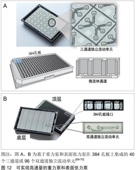
[69] VAN DV, ZHU D, RAMAKERS C, et al.Perfused 3D angiogenic sprouting in a high-throughput in vitro platform. Angiogenesis. 2019;22(1):157-165.
[70] VAN DUINEN V, VAN D HA, TRIETSCH SJ, et al. 96 perfusable blood vessels to study vascular permeability in vitro. Sci Rep-UK.2017;7(1):18071.
|