中国组织工程研究 ›› 2017, Vol. 21 ›› Issue (32): 5158-5163.doi: 10.3969/j.issn.2095-4344.2017.32.013
• 皮肤粘膜组织构建 skin and mucosal tissue construction • 上一篇 下一篇
瘢痕成熟过程中血管生成素1表达与瘢痕血管的变化
吴子涵1,李高峰2
- (1吉首大学第一附属医院湘西自治州人民医院,湖南省吉首市 416000;2湖南师范大学第一附属医院湖南省人民医院,湖南省长沙市 410000)
Correlation of angiopoietin-1 with angiogenesis during scar formation
Wu Zi-han1, Li Gao-feng2
- (1People’s Hospital of Xiangxi Autonomous Prefecture, the First Affiliated Hospital of Jishou University, Jishou 416000, Hunan Province, China; 2Hunan Provincial People’s Hospital, the First Affiliated Hospital of Hunan Normal University, Changsha 41000, Hunan Province, China)
摘要:
文章快速阅读:
.jpg) 文题释义:
血管生成:从已有的毛细血管或毛细血管后静脉发展而形成新的血管,主要包括:激活期血管基底膜降解,血管内皮细胞的激活、增殖、迁移,重建形成新的血管和血管网,是一个涉及多种细胞因子的复杂过程。
血管生成素1:为重要的血管生成因子,能保证新生血管内皮细胞的正确组装、促进内皮周围支持细胞的聚集和血管重塑,从而维持新生血管的完整性和稳定性,对新生血管的成熟具有重要作用。
文题释义:
血管生成:从已有的毛细血管或毛细血管后静脉发展而形成新的血管,主要包括:激活期血管基底膜降解,血管内皮细胞的激活、增殖、迁移,重建形成新的血管和血管网,是一个涉及多种细胞因子的复杂过程。
血管生成素1:为重要的血管生成因子,能保证新生血管内皮细胞的正确组装、促进内皮周围支持细胞的聚集和血管重塑,从而维持新生血管的完整性和稳定性,对新生血管的成熟具有重要作用。
文题释义:
血管生成:从已有的毛细血管或毛细血管后静脉发展而形成新的血管,主要包括:激活期血管基底膜降解,血管内皮细胞的激活、增殖、迁移,重建形成新的血管和血管网,是一个涉及多种细胞因子的复杂过程。
血管生成素1:为重要的血管生成因子,能保证新生血管内皮细胞的正确组装、促进内皮周围支持细胞的聚集和血管重塑,从而维持新生血管的完整性和稳定性,对新生血管的成熟具有重要作用。摘要
背景:目前关于血管内皮细胞生长因子与瘢痕的研究较多,而血管生成素1与瘢痕的研究少有报道。
目的:分析瘢痕成熟过程中血管生成素1的表达与瘢痕血管变化的关系。
方法:取雌雄不限的新西兰大白兔,在耳腹侧中部相同位置制造瘢痕模型,分别在上皮化后1,2,4,8,12周切取兔耳瘢痕组织标本及兔耳腹侧正常皮肤组织,采用苏木精-伊红染色观察瘢痕成熟过程中的大体形态,CD34免疫组织化学染色观察瘢痕血管变化,Western-blot检测血管生成素1表达。
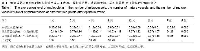
结果与结论:①瘢痕中血管生成素1表达的趋势是先升高,至上皮化后2周最高,之后逐渐降低,至上皮化后12周最小,接近正常皮肤血管生成素1的表达;②微血管总数在上皮化后4周最高,以后逐渐降低;③成熟血管数呈逐渐升高的趋势;④成熟血管数/微血管总数比值呈逐渐升高的趋势;⑤兔耳瘢痕萎缩成熟过程中血管生成素1表达与成熟血管数呈负相关(P < 0.05),血管生成素1表达与成熟血管数/微血管总数比值呈负相关(P < 0.05);⑥血管生成素1在瘢痕萎缩成熟过程中对瘢痕血管成熟可能起重要作用。
中国组织工程研究杂志出版内容重点:组织构建;骨细胞;软骨细胞;细胞培养;成纤维细胞;血管内皮细胞;骨质疏松;组织工程
ORCID: 0000-0002-2326-0388(吴子涵)
中图分类号:


.jpg) 文题释义:
血管生成:从已有的毛细血管或毛细血管后静脉发展而形成新的血管,主要包括:激活期血管基底膜降解,血管内皮细胞的激活、增殖、迁移,重建形成新的血管和血管网,是一个涉及多种细胞因子的复杂过程。
血管生成素1:为重要的血管生成因子,能保证新生血管内皮细胞的正确组装、促进内皮周围支持细胞的聚集和血管重塑,从而维持新生血管的完整性和稳定性,对新生血管的成熟具有重要作用。
文题释义:
血管生成:从已有的毛细血管或毛细血管后静脉发展而形成新的血管,主要包括:激活期血管基底膜降解,血管内皮细胞的激活、增殖、迁移,重建形成新的血管和血管网,是一个涉及多种细胞因子的复杂过程。
血管生成素1:为重要的血管生成因子,能保证新生血管内皮细胞的正确组装、促进内皮周围支持细胞的聚集和血管重塑,从而维持新生血管的完整性和稳定性,对新生血管的成熟具有重要作用。