中国组织工程研究 ›› 2017, Vol. 21 ›› Issue (26): 4228-4233.doi: 10.3969/j.issn.2095-4344.2017.26.022

• 生物材料临床实践 clinical practice of biomaterials • 上一篇    下一篇

基于CT灌注评价早期钛网修补颅骨缺损对脑血流量及神经功能康复的影响:随机对照临床试验

庄亚瑟,方志成,刘伯毅,陈  黎,俞旦峰
  

  1. 十堰市太和医院(湖北医药学院附属医院)重症医学科,湖北省十堰市  442000
  • 收稿日期:2017-05-25 出版日期:2017-09-18 发布日期:2017-09-28
  • 通讯作者: 方志成,硕士,主任医师,十堰市太和医院(湖北医药学院附属医院)重症医学科,湖北省十堰市 442000
  • 作者简介:庄亚瑟,男,1990年生,湖北省丹江口市人,汉族,宁夏医科大学毕业,硕士,医师,主要擅长于脑外伤等疾病的急危重症治疗。
  • 基金资助:
    湖北省卫生计生科研项目(WJ2015MB308)

Effects of early skull repair with titanium mesh on cerebral blood flow and neurological recovery: a randomized controlled clinical trial based on CT perfusion evaluation

Zhuang Ya-se, Fang Zhi-cheng, Liu Bo-yi, Chen Li, Yu Dan-feng
  

  1. Department of Critical Care Medicine, Taihe Hopsital (Affiliated Hospital of Hubei University of Medicine), Shiyan 442000, Hubei Province, China
  • Received:2017-05-25 Online:2017-09-18 Published:2017-09-28
  • Contact: Fang Zhi-cheng, Master, Chief physician, Department of Critical Care Medicine, Taihe Hopsital (Affiliated Hospital of Hubei University of Medicine), Shiyan 442000, Hubei Province, China
  • About author:Zhuang Ya-se, Master, Physician, Department of Critical Care Medicine, Taihe Hopsital (Affiliated Hospital of Hubei University of Medicine), Shiyan 442000, Hubei Province, China
  • Supported by:
    the Scientific Research Project for Health and Family Planning in Hubei Province, No. WJ2015MB308

摘要:

文章快速阅读:


文题释义:
CT 灌注技术:是CT检查的附加功能,是通过螺旋CT仪器来实现的一项技术,它基于核医学的放射性示踪剂稀释和中心容积定律原理,具有多参数、扫描迅速、设备普及等优点。
CT 灌注评价脑血流量:借助CT灌注技术观测对比剂在颅脑的浓度及分布是否随时间变化的情况,评估脑组织血液动力学情况。首先静脉注射对比剂,当其通过脑组织和血管时,扫描选定区域获得密度曲线,时间-密度曲线反映的是测定区域中对比剂浓度和时间的相关性,间接反映脑组织血液灌注量。
 
背景:创伤性脑损伤后去骨瓣减压治疗形成了颅骨缺损,目前公认最合适的颅骨修补人工材料为金属钛网,其生物相容性良好,在临床上获得了广泛应用,但脑损伤后颅骨缺损修复的时机仍存在争议。
目的:采用CT 灌注技术比较早期、晚期钛网修补颅骨缺损前后患者的脑组织血流灌注变化以及神经功能康复情况。
方法:试验为单中心、前瞻性、观察性临床试验,在中国湖北省,湖北医药学院附属太和医院完成。选取2013年1月至2016年1月收治的颅脑损伤后去骨瓣减压患者86例,均采用钛网进行颅骨修复,随机分为2组,试验组40例行早期颅骨修补(去骨瓣减压术后1-3个月),对照组46例行晚期颅骨修补(去骨瓣减压术后6-12个月)。颅骨修补术前3 d和术后第10天,应用CT灌注技术观察脑组织血液灌注量变化;术后30 d评价日常活动能力Barthel 指数。试验于2017年7月12日在北美临床试验中心注册(NCT03222297)。试验方案经十堰市太和医院伦理委员会批准,批准号为2012第(08)号。临床试验研究的实施符合《赫尔辛基宣言》和医院对人体研究的相关伦理要求。参与试验的患病个体及其家属为自愿参加,均对试验过程完全知情同意,在充分了解治疗方案的前提下签署“知情同意书”。
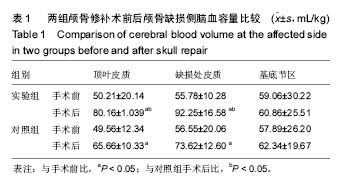
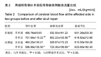
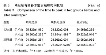
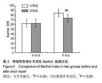
结果与结论:①两组患者颅骨修补后缺损侧顶叶皮质、缺损处皮质的脑血容量和脑血流量均高于颅骨修补前(P < 0.05),达峰时间则低于颅骨修补前(P < 0.05);颅骨修补后试验组的缺损侧顶叶皮质、缺损处皮质的脑血容量和脑血流量均高于对照组(P < 0.05),达峰时间则低于对照组(P < 0.05);②颅骨修补术后30 d试验组的Barthel 指数得分高于对照组(P < 0.05);③说明早期行颅骨缺损修补,对改善患侧脑组织灌注情况有利,进而对患侧神经功能的恢复有意义,而CT灌注观测脑血流动力学的变化安全有效。

关键词: 生物材料, 材料相容性, 颅骨缺损, 钛网, CT灌注, 脑血流量, Barthel 指数

Abstract:

BACKGROUND: A skull defect is inevitable after decompression treatment for traumatic brain injury. Titanium mesh as the most recognized skull repair material has good biocompatibility and has been widely used in clinical practice. However, the timing for skull repair after brain injury is still in dispute.
OBJECTIVE: To compare the changes of brain perfusion and the recovery of neurological function in patients with skull defects before and after early and late-stage titanium mesh repair based on CT perfusion technique.
METHODS: This was a single-center, prospective, observational clinical trial that was completed at the Taihe Hospital, Hubei University of Medicine in Hubei Province, China. Eighty-six patients with craniocerebral injury who had undergone decompression with removal of bone flap from January 2013 to January 2016 were recruited and subjected to skull repair using titanium mesh. All the patients were randomized into two groups: test group (n=40) with early skull repair within 1-3 months after decompression and control group (n=46) with late-stage skull repair within 6-12 months after decompression. CT perfusion technology was used to observe changes of brain perfusion at 3 days operatively and at 10 days postoperatively. The Barthel index was evaluated at 30 days postoperatively. The trial was registered with ClinicalTrial.gov (identifier: NCT03222297) on July 12th, 2017. The study protocol was approved by the Ethics Committee of Taihe Hospital with the approval No. 2012 (08), and performed in accordance with the Declaration of Helsinki, formulated by the World Health Organization and the hospital's ethical requirements for human research. All the patients and their families were voluntary to participate in the trial, were fully informed of the trial process, and then signed the informed consent prior to the initialization of the trial.
RESULTS AND CONCLUSION: The postoperative cerebral blood volume and cerebral blood flow at the parietal cortex on the side of skull defect and at the cortex in the defect region were significantly higher in the two group than the baseline (P < 0.05), while the time to peak was lower than the baseline (P < 0.05). Compared with the control group, significantly higher cerebral blood volume and cerebral blood flow as well as shorter time to peak were observed in the test group (P < 0.05). The Barthel index of the test group was also significantly higher than that of the control group at 30 days postoepratively (P < 0.05). Overall, early skull repair with titanium mesh is helpful to improve the cerebral blood perfusion at the affected side and the recovery of neurological function. In addition, CT perfusion technology is a safe and effective method to monitor hemodynamic changes in the brain.

Key words: Biocompatible Materials, Decompressive Craniectomy, Titanium, Tissue Engineering

中图分类号: