中国组织工程研究 ›› 2017, Vol. 21 ›› Issue (26): 4119-4124.doi: 10.3969/j.issn.2095-4344.2017.26.004

• 组织工程骨及软骨材料 tissue-engineered bone and cartilage materials • 上一篇    下一篇

β磷酸三钙和α半水硫酸钙复合人工骨生物相容性及在脊柱融合模型中的应用

谭海涛,孟志斌,李  俊,黄  涛,王挺锐,符国良
  

  1. 海南医学院第一附属医院骨科,海南省海口市  570102
  • 收稿日期:2017-08-02 出版日期:2017-09-18 发布日期:2017-09-28
  • 通讯作者: 孟志斌,硕士,教授,主任医师,海南医学院第一附属医院骨科,海南省海口市 570102
  • 作者简介:谭海涛,男,汉族,1982年生,湖南省常德市人,硕士,主治医师。
  • 基金资助:
    海南省自然科学基金面上项目(20168297)

Biocompatibility of beta-tricalcium phosphate/alpha-calcium sulfate hemihydrate artificial bone and its application in a spinal fusion model

Tan Hai-tao, Meng Zhi-bin, Li Jun, Huang Tao, Wang Ting-rui, Fu Guo-liang
  

  1. Department of Orthopaedics, the First Affiliated Hospital of Hainan Medical University, Haikou 570102, Hainan Province, China
  • Received:2017-08-02 Online:2017-09-18 Published:2017-09-28
  • Contact: Meng Zhi-bin, Master, Professor, Chief physician, Department of Orthopaedics, the First Affiliated Hospital of Hainan Medical University, Haikou 570102, Hainan Province, China
  • About author:Tan Hai-tao, Master, Attending physician, Department of Orthopaedics, the First Affiliated Hospital of Hainan Medical University, Haikou 570102, Hainan Province, China
  • Supported by:
     the Natural Science Foundation of Hainan Province, No. 20168297

摘要:

文章快速阅读:


文题释义:
β磷酸三钙:磷酸钙类人工骨中常用材料,通过不同的制备工艺或改变材料的孔隙结构,获得不同的降解性能。
磷酸钙类材料:具有优良的理化性质和生物学特性,能保留天然松质骨的孔隙结构,为新生骨组织和血管的长入提供理想的通道。
 
背景:β磷酸三钙和α半水硫酸钙备的人工复合骨具有多孔颗粒形态,生物相容性较高,用于脊柱融合模型中有助于融合率的提高,但该结论尚未得到证实。
目的:观察β磷酸三钙/α半水硫酸钙复合人工骨的制备方法、生物相容性及在脊柱融合模型中的应用效果。
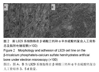
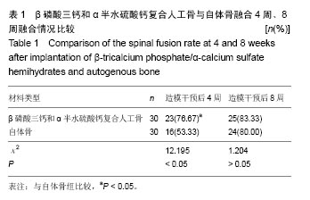
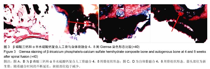
方法:①将二水硫酸钙在定点的条件、温度下脱水制备α半水硫酸钙;取健康牛松质骨经脱细胞、脱脂后在特点的条件、温度下煅烧制备β磷酸三钙多孔颗粒,溶于无水乙醇中,混悬,烘干后制备β磷酸三钙和α半水硫酸钙复合人工骨。取购置的兔骨膜成骨细胞与复合材料共同培养,观察细胞形态、黏附能力及增殖活力;②取新西兰大白兔30只,建立双侧新西兰大白兔胸椎多节段后外侧脊柱融合模型,左侧植入β磷酸三钙和α半水硫酸钙复合人工骨,右侧植入自体骨进行对照,比较2组融合率变化。
结果与结论:①细胞形态:相差显微镜观察可见,L929系细胞株培养3 d后在β磷酸三钙和α半水硫酸钙复合人工骨上贴壁数量相对较少;培养5 d后细胞在人工骨上贴壁相对密集;扫描电镜下β磷酸三钙和α半水硫酸钙复合人工骨表面存在许多结晶颗粒,表面吸附细胞相对较多;②β磷酸三钙和α半水硫酸钙复合人工骨植入后4周融合率,高于自体骨(P < 0.05);③自体骨植入后4周组织切片中骨侧小梁稀疏杂乱,可见新生的骨组织,无核的自体骨移植骨块占据主导地位;融合后8周自体骨移植骨周围新生骨组织进一步增多;β磷酸三钙和α半水硫酸钙复合人工骨植入后4周材料结构清晰可见,未见降解碎片,内部存在少许新生骨组织;植入后8周材料被新生骨组织包围,骨小梁增粗,人工骨开始降解;④结果证实:制备的β磷酸三钙和α半水硫酸钙复合人工骨用于脊柱融合模型中能获得较高的融合率。

关键词: 生物材料, 骨生物材料, β磷酸三钙, α半水硫酸钙, 复合人工骨, 制备方法, 生物相容性, 脊柱融合模型, 增殖活力, 海南省自然科学基金

Abstract:

BACKGROUND: Beta-tricalcium phosphate (β-TCP)/alpha-calcium sulfate hemihydrate (α-CSH) artificial composite bone has a porous morphology and good biocompatibility, and it is helpful to improve the fusion rate in a spinal fusion model, which however has not yet been confirmed.
OBJECTIVE: To investigate the preparation methods, biocompatibility and application effect of β-TCP/α-CSH composite bone in the spinal fusion model.
METHODS: (1) Calcium sulfate dihydrate under certain conditions and at a proper temperature can be dehydrated to prepare α-CSH. Healthy bovine cancellous bone was decellularized, degreased and sintered under the certain condition and at the certain temperature to prepare β-TCP particles. Then, the β-TCP particles were dissolved in anhydrous ethanol, suspended, dried, and then used to prepare the β-TCP/α-CSH composite bone. Osteoblasts from the rabbit periosteum were co-cultured with the composite bone, and then cell morphology, adhesion and proliferation were observed. (2) Twenty New Zealand white rabbits were selected to make bilaterally posterolateral spinal fusion models of the multiple thoracic vertebrae, in which β-TCP/α-CSH composite bone was implanted into the left side (experimental group) and autogenous bone implanted into the right side (control group). The spinal fusion rate was compared between the two groups.
RESULTS AND CONCLUSION: (1) Under the phase contrast microscope, a relatively small amount of L929 cells adhered to the composite bone after 3 days of co-culture, while the number of adherent cells became relatively dense. Under the scanning electron microscope, there were many crystalline particles on the surface of the composite bone, indicating a higher number of adherent cells on the composite bone surface. (2) The spinal fusion rate was increased at 4 weeks after implantation of β-TCP/α-CSH composite bone, which was significantly higher than that after implantation of autogenous bone (P < 0.05). (3) At 4 weeks after autogenous bone implantation, the bone trabecular bone was scarce and clumped, and newborn bone tissues and the boneless autograft bone were dominant. After 8 weeks after spinal fusion, the newborn bone tissues around the autograft were further increased. At 4 weeks after β-TCP/α-CSH composite bone implantation, there was no degradation of debris, but existed a few new bone tissues; at 8 weeks after implantation, the composite bone was surrounded by newborn bone tissues, and thickened trabecular bone and degradation of the composite bone were found. To conclude, the prepared β-TCP/α-CSH composite bone can achieve a higher fusion rate in the spinal fusion model.

Key words: Tissue Engineering, Biocompatible Materials, Spine

中图分类号: