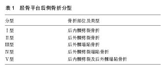
| [1] Bhattacharyya T, McCarty LP 3rd, Harris MB, et al. The posterior shearing tibial plateau fracture: treatment and results via a posterior approach. J Orthop Trauma. 2005; 19:305-310.[2] Chang SM,Zheng HP,Li HF,et al.Treatment of isolated posterior coronal fracture of the lateral tibial plateau through posterolateral approach for direct exposure and buttress plate fixation. Arch Orthop Trauma Surg. 2009; 129(7): 955-962.[3] Higgins TF,Klatt J,Bachus KN.Biomechanical analysis of bicondylar tibial plateau fixation: How does lateral locking plate fixation compare to dual plate fixation.J Orthop Trauma. 2007; 21(5): 30l-306.[4] Zhu Y, Yang G, Luo CF,et al. Computed tomography-based Three-Column Classification in tibial plateau fractures: introduction of its utility and assessment of its reproducibility. J Trauma Acute Care Surg. 2012; 73(3): 731-737.[5] Heidari N,Lidder S,Grechenig W,et al. The risk of injury to the anterior tibial artery in the posterolateral approach to the tibia plateau:acadaver study.J Orthop Trauma. 2013;27(4): 221-225.[6] Sun H,Luo CF,Yang G,et al.Anatomical evaluation of the modified posterolateral approach for posterolateral tibial plateau fracture.Eur J Orthop Surg Traumatol. 2013; 23(7): 809-818.[7] 胡孙君,张世民,张英琪,等. 胫骨平台后外侧象限骨折手术入路的深层解剖及后外侧与后内侧比较[J]. 中国临床解剖学杂志, 2015,33(5): 497-501.[8] Pacheco RJ,Ayre CA,Bollen SR.Posterolateral comer injuries of the knee:a serious injury commonly missed.J Bone Joint Surg Br. 2011; 93(2): 194-197.[9] Khan RM,Khan SH,Ahmad AJ,et al. Tibial plateau fractures. A new classification scheme. Clin Orthop Relat Res. 2000; 375: 231-242.[10] Luo CF,Sun H,Zhang B,et al.Three-column fixation for complex tibial plateau fractures.J Orthop Trauma. 2010; 24(11): 683-692.[11] Chen HW,Chen CQ,Yi XH. Posterior tibial plateau fracture: a new treatment-oriented classification and surgical management. Int J Clin Exp Med. 2015; 8(1): 472-479.[12] 陈红卫,赵钢生,王子阳,等. 胫骨平台后髁骨折的CT分型[J]. 中华医学杂志.2011; 91(3): 180-184.[13] 高翔,李杭,郑强,等.胫骨后外侧平台骨折的CT形态学研究[J]. 中华骨科杂志. 2014;34(7)709-716.[14] Barei DP,O’Mara Tj,Taitsman LA,et al.Frequency and fracture morphology of the posteromedial fragment in bicondylar tibial plateau fracture patterns.J Orthop Trauma. 2008; 22(3): 176-182.[15] Sohn HS,Yoon YC,Cho JW,et al.Incidence and fracture morphology of posterolateral fragments in lateral and bicondylar tibial plateaufractures.J Orthop Trauma. 2015; 29(2):91-97.[16] Zhu Y,Meili S,Dong MJ,et al. Pathoanatomy and incidence of the posterolateral fractures in bicondylar tibial plateau fractures: a clinical computed tomography-based measurement and the associated biomechanical model simulation. Arch Orthop Trauma Surg. 2014; 134(10): 1369-1380.[17] Xiang G,Zhi-Jun P,Qiang Z,et al.Morphological characteristics of posterolateral articular Fragments in tibial plateau fractures.Orthopedics. 2013; 36(10): e1256-e1261.[18] Zhai Q,Luo C,Zhu Y,et al.Morphological characteristics of split-depression fractures of the lateral tibial plateau(Schatzker type II):a computer-tomography-based study.Int Orthop. 2013; 37(5): 911-917.[19] Kenneth AE. Split depression posterolateral tibial plateau fracture direct open reduction and internal fixation. Tech Knee Surg. 2005; 4(4): 257-262.[20] 刘立峰,蔡锦力,梁进. 胫骨平台后外壁骨折的治疗[J]. 中国骨伤,2003,16(6): 338-339.[21] Hsieh CH. Treatment of the posterolateral tibial plateau fractures using the anterior surgical approach.J Biomed Sci. 2010; 6(4): 316-320.[22] 冯刚,潘志军,李杭,等. 双锁定钢板交叉支撑固定治疗累及后外侧的C3型胫骨平台骨折[J]. 中华骨折杂志,2014; 34(7): 695-702.[23] Sassoon AA,Torchia Me,Cross Ww,et al.Fibular shaft allograft support of posterior joint depression in tibial plateau fractures.J Orthop Trauma. 2014; 28(7): e169-e175.[24] Solomon LB,Stevenson AW,Baird RP,et al.Posterolateral transfibular approach to tibial plateau fractures:technique,results,and rationale.J Orthop Trauma. 2010; 24(8): 505-514.[25] Yu B,Han K,Zhan C,et al.Fibular head osteotomy:a new approach for the treatment of lateral or posterolateral tibial plateau fractures.Knee. 2010; 17(5): 313-318.[26] 庄岩,王鹏飞,张堃,等. 经腓骨截骨入路治疗胫骨平台后外侧骨折的疗效观察. 中华骨科杂志. 2012; 32(8): 732-738.[27] Zhang W,Luo CF,Putnis S,et al.Biomechanical analysis of four different fixations for the posterolateral shearing tibial plateau fracture. Knee. 2012; 19(2): 94-98.[28] Carlson DA.Posterior bicondylar tibial plateau fractures.J Orthop Trauma. 2005; 19(2): 73-78.[29] Tao J,Hang DH,Wang QG,et al.The posterolateral shearing tibial plateau fracture: treatment and results via a modified posterolateral approach.Knee. 2008; 15(6): 473-479.[30] Liu GY,Xiao BP,Luo CF, et al.Results of a modified posterolateral approach for the isolated posterolateral tibial plateau fracture. Indian J Orthop. 2016; 50(2):117-122.[31] Frosch KH,Balcarek P,Walde T,et al.A new posterolateral approach without fibula osteotomy for the treatment of tibial plateau fractures.J Orthop Trauma. 2010; 24(8): 515-520.[32] Chen HW,Luo CF. Extended anterolateral approach for treatment of posterolateral tibial plateau fractures improves operative procedure and patient prognosis. Int J Clin Exp Med. 2015; 8(8): 13708-13715.[33] 朱海涛,王文跃,王俭,等. 外后侧弧形切口双肌间隙入路治疗胫骨后外侧平台塌陷骨折[J]. 中华骨科杂志, 2014; 34(7): 703-708.[34] Chen HW, Zhou SH, Liu GD,et al. An extended anterolateral approach for posterolateral tibial plateau fractures. Knee Surg Sports Traumatol Arthrosc. 2015; 23(12):3750-3755.[35] Hu SJ,Chang SM,Zhang YQ,et al.The anterolateral supra-fibular-head approach for plating posterolateral tibial plateau fractures: A novel surgical technique. Injury. 2016; 47(2): 502-507.[36] 刘观燚,罗从风,赵华国,等. 改良后外侧入路治疗孤立性后外侧胫骨平台骨折[J]. 中华创伤骨科杂志,2015,17(10): 895-898.[37] He X,Ye P,Hu Y,et al.A posterior inverted L-shaped approach for the treatment of posterior bicondylar tibial plateau fractures.Arch Orthop Trauma Surg. 2013;133(1): 23-28.[38] Solomon LB,Stevenson AW,Lee YC,et al.Posterolateral and antero-lateral approaches to unicondylar posterolateral tibial plateau fractures:a comparative study.Injury. 2013; 44(11): 1561-1568.[39] Qiu WJ,Zhan Y,Sun H,et al. A posterior reversed L-shaped approach for the tibial plateau fractures—A prospective study of complications (95 cases) . Injury. 2015; 46: 1613–1618.[40] Berber R,Lewis CP,Copas D,et al.Postero-medial approach for complex tibial plateau injuries with a postero-medial or postero-lateral shear fragment. Injury. 2014; 45(4): 757-765.[41] Sun H,Zhai QL,Xu YF,et al.Combined approaches for fixation of Schatzker type II tibial plateau fractures involving the posterolateralcolumn: a prospective observational cohort study. Arch Orthop Trauma Surg. 2015; 135(2): 209-221.[42] Chang SM,Hu SJ,Zhang YQ,et al.A surgical protocol for bicondylar four-quadrant tibial plateau fractures. Int Orthop. 2014;38(12): 2559-2564.[43] Jiang R,Luo CF,Zeng BF.Biomechanical evaluation of different fixation methods for fracture dislocation involving the proximal tibia.Clin Biomech(Bristol,Avon). 2008; 23(8): 1059-1064.[44] Zeng ZM,Luo CF,Putnis S,et al.Biomechanical analysis of posteromedial tibial plateau split fracture fixation.Knee. 2011; 18(1): 51-54.[45] 张巍,罗从风,曾炳芳.四种不同内固定治疗胫骨平台后外侧剪应力骨折的生物力学研究[J].中华创伤骨科杂志. 2010; 12(11): 1069-1073. |
.jpg)
.jpg)