| [1]Zhou Y, Wu C, Xiao Y. The stimulation of proliferation and differentiation of periodontal ligament cells by the ionic products from Ca7Si2P2O16 bioceramics. Acta Biomater. 2012;8(6):2307-2316.[2]Zhang Y, Fan W, Ma Z, et al. The effects of pore architecture in silk fibroin scaffolds on the growth and differentiation of mesenchymal stem cells expressing BMP7. Acta Biomater. 2010;6(8):3021-3028.[3]Cho SY, Park HH, Jin HJ. Controlling pore size of electrospun silk fibroin scaffold for tissue engineering. Polym Korea. 2012; 36(5):651-655.[4]Hench LL, Wilson J. Surface-active biomaterials. Science. 1984;226(4675):630-636.[5]Zhu M, Shi JL, He QJ, et al. An emulsification-solvent evaporation route to mesoporous bioactive glass microspheres for bisphosphonate drug delivery. J Mater Sci. 2012; 47(5):2256-2263.[6]Wang D, Lin H, Jiang J, et al. One-pot synthesis of magnetic, macro/mesoporous bioactive glasses for bone tissue engineering. Sci Technol Adv Mater. 2013;14(2):025004.[7]Baino F, Fiorilli S, Mortera R, et al. Mesoporous bioactive glass as a multifunctional system for bone regeneration and controlled drug release. J Appl Biomater Funct Mater. 2012;10(1):12-21.[8]Wu C, Chang J. Mesoporous bioactive glasses: structure characteristics, drug/growth factor delivery and bone regeneration application. Interface Focus. 2012;2(3):292-306.[9]Wang X, Chen W, Liu Q, et al. Function and mechanism of mesoporous bioactive glass adsorbed epidermal growth factor for accelerating bone tissue regeneration. Biomed Mater. 2017;12(2):025020.[10]Shoaib M, Saeed A, Akhtar J, et al. Potassium-doped mesoporous bioactive glass: Synthesis, characterization and evaluation of biomedical properties. Mater Sci Eng C Mater Biol Appl. 2017;75:836-844. [11]Qi X, Pei P, Zhu M, et al. Three dimensional printing of calcium sulfate and mesoporous bioactive glass scaffolds for improving bone regeneration in vitro and in vivo. Sci Rep. 2017;7:42556.[12]Schumacher M, Reither L, Thomas J, et al. Calcium phosphate bone cement/mesoporous bioactive glass composites for controlled growth factor delivery. Biomater Sci. 2017;5(3):578-588. [13]Zhang Q, Chen X, Geng S, et al. Nanogel-based scaffolds fabricated for bone regeneration with mesoporous bioactive glass and strontium: In vitro and in vivo characterization. J Biomed Mater Res A. 2017;105(4):1175-1183. [14]Wang X, Li W. Biodegradable mesoporous bioactive glass nanospheres for drug delivery and bone tissue regeneration. Nanotechnology. 2016;27(22):225102. [15]Wu C, Xia L, Han P, et al. Europium-Containing Mesoporous Bioactive Glass Scaffolds for Stimulating in Vitro and in Vivo Osteogenesis. ACS Appl Mater Interfaces. 2016;8(18): 11342-11354.[16]Marie PJ, Ammann P, Boivin G, et al. Mechanisms of action and therapeutic potential of strontium in bone. Calcif Tissue Int. 2001;69(3):121-129.[17]Gentleman E, Fredholm YC, Jell G, et al. The effects of strontium-substituted bioactive glasses on osteoblasts and osteoclasts in vitro. Biomaterials. 2010;31(14):3949-3956.[18]Zreiqat H, Ramaswamy Y, Wu C, et al. The incorporation of strontium and zinc into a calcium-silicon ceramic for bone tissue engineering. Biomaterials. 2010;31(12):3175-3184.[19]Tsang VL, Bhatia SN. Three-dimensional tissue fabrication. Adv Drug Deliv Rev. 2004;56(11):1635-1647.[20]Rao RB, Krafcik KL, Morales AM, et al. Microfabricated Deposition Nozzles for Direct-Write Assembly of Three-Dimensional Periodic Structures. Advanced Materials. 2005; 17(3):289-293.[21]Franco J, Hunger P, Launey ME, et al. Direct write assembly of calcium phosphate scaffolds using a water-based hydrogel. Acta Biomater. 2010;6(1):218-228.[22]Khan MN, Islam JM, Khan MA. Fabrication and characterization of gelatin-based biocompatible porous composite scaffold for bone tissue engineering. J Biomed Mater Res A. 2012;100(11):3020-3028.[23]Wang Z, Sakakibara T, Sudo A, et al. Porosity of β-tricalcium phosphate affects the results of lumbar posterolateral fusion. J Spinal Disord Tech. 2013;26(2):E40-45.[24]Yan X, Huang X, Yu C, et al. The in-vitro bioactivity of mesoporous bioactive glasses. Biomaterials. 2006;27(18):3396-3403.[25]Takeuchi A, Mishina Y, Miyaishi O, et al. Heterozygosity with respect to Zfp148 causes complete loss of fetal germ cells during mouse embryogenesis. Nat Genet. 2003;33(2): 172-176.[26]Melchels FP, Barradas AM, van Blitterswijk CA, et al. Effects of the architecture of tissue engineering scaffolds on cell seeding and culturing. Acta Biomater. 2010;6(11):4208-4217.[27]Yun HS, Park JW, Kim SH, et al. Effect of the pore structure of bioactive glass balls on biocompatibility in vitro and in vivo. Acta Biomater. 2011;7(6):2651-2660.[28]Wang C, Zhao Q, Wang M, et al. Cryogenic 3D printing for producing hierarchical porous and rhBMP-2-loaded Ca-P/PLLA nanocomposite scaffolds for bone tissue engineering. Biofabrication. 2017;9(2):025031. [29]Sears N, Dhavalikar P, Whitely M,et al. Fabrication of biomimetic bone grafts with multi-material 3D printing. Biofabrication. 2017;9(2):025020.[30]Wang Y, Wang K, Li X, et al. 3D fabrication and characterization of phosphoric acid scaffold with a HA/β-TCP weight ratio of 60:40 for bone tissue engineering applications. PLoS One. 2017;12(4):e0174870.[31]Shao H, Ke X, Liu A,et al. Bone regeneration in 3D printing bioactive ceramic scaffolds with improved tissue/material interface pore architecture in thin-wall bone defect. Biofabrication. 2017;9(2):025003.[32]Qi X, Pei P, Zhu M, et al.Three dimensional printing of calcium sulfate and mesoporous bioactive glass scaffolds for improving bone regeneration in vitro and in vivo. Sci Rep. 2017;7:42556. [33]Bendtsen ST, Quinnell SP, Wei M. Development of a novel alginate-polyvinyl alcohol-hydroxyapatite hydrogel for 3D bioprinting bone tissue engineered scaffolds. J Biomed Mater Res A. 2017;105(5):1457-1468.[34]Egorov AA, Fedotov AY, Mironov AV, et al. 3D printing of mineral-polymer bone substitutes based on sodium alginate and calcium phosphate. Beilstein J Nanotechnol. 2016;7: 1794-1799. [35]Li C, Jiang C, Deng Y, et al. RhBMP-2 loaded 3D-printed mesoporous silica/calcium phosphate cement porous scaffolds with enhanced vascularization and osteogenesis properties. Sci Rep. 2017;7:41331.[36]Causa F, Battista E, Della Moglie R, et al. Surface investigation on biomimetic materials to control cell adhesion: the case of RGD conjugation on PCL. Langmuir. 2010;26(12): 9875-9884.[37]Huang ZH, Song B, Chen YF, et al. Effect of polycaprolactone-ascobic acid scaffold in repairing articular cartilage defects in rabbits. Nan Fang Yi Ke Da Xue Xue Bao. 2017;37(5):607-613.[38]Esmaeilzadeh J, Hesaraki S, Hadavi SM, et al. Poly (d/l) lactide/polycaprolactone/bioactive glasss nanocomposites materials for anterior cruciate ligament reconstruction screws: The effect of glass surface functionalization on mechanical properties and cell behaviors. Mater Sci Eng C Mater Biol Appl. 2017;77:978-989. [39]Kong J, Yu Y, Pei X, et al.Polycaprolactone nanocomposite reinforced by bioresource starch-based nanoparticles. Int J Biol Macromol. 2017;102:1304-1311.[40]Gomes S, Rodrigues G, Martins G, et al. Evaluation of nanofibrous scaffolds obtained from blends of chitosan, gelatin and polycaprolactone for skin tissue engineering. Int J Biol Macromol. 2017;102:1174-1185. [41]Shim JH, Won JY, Park JH, et al. Effects of 3D-Printed Polycaprolactone/β-Tricalcium Phosphate Membranes on Guided Bone Regeneration. Int J Mol Sci. 2017;18(5): E899.[42]Wu C, Ramaswamy Y, Kwik D, et al. The effect of strontium incorporation into CaSiO3 ceramics on their physical and biological properties. Biomaterials. 2007;28(21):3171-3181.[43]Zhang J, Zhao S, Zhu Y, et al. Three-dimensional printing of strontium-containing mesoporous bioactive glass scaffolds for bone regeneration. Acta Biomater. 2014;10(5):2269-2281.[44]Valerio P, Pereira MM, Goes AM, et al. The effect of ionic products from bioactive glass dissolution on osteoblast proliferation and collagen production. Biomaterials. 2004; 25(15):2941-2948. |
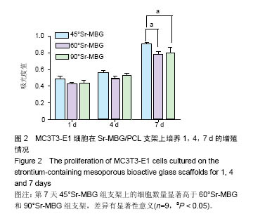
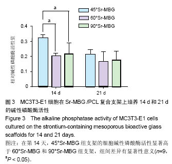
.jpg)
.jpg)
.jpg)
.jpg)