中国组织工程研究 ›› 2017, Vol. 21 ›› Issue (18): 2789-2795.doi: 10.3969/j.issn.2095-4344.2017.18.001

• 组织工程骨及软骨材料 tissue-engineered bone and cartilage materials •    下一篇

新型骨植入材料多孔钽-骨结合界面成骨以及相关成骨因子的表达及意义

赖振权1,崔逸爽1,陈  超2,周国龙1,潘祥宇1,王  茜3,甘洪全4,王志强4,李琪佳1
  

  1. 华北理工大学,1医学实验中心,3基础医学院,河北省唐山市  063000;2河北省遵化市人民医院病理科,河北省遵化市  064200;4华北理工大学附属医院骨科,河北省唐山市  063000
  • 收稿日期:2017-02-07 出版日期:2017-06-28 发布日期:2017-07-07
  • 通讯作者: 李琪佳,教授,硕士生导师,华北理工大学医学实验中心,河北省唐山市 063000
  • 作者简介:赖振权,男,1993年生,福建省泉州市人,汉族,2017年华北理工大学毕业,主要从事骨组织工程研究。
  • 基金资助:
    国家科技部科技支撑课题(2012BAE06B03);河北省科技支撑课题(16277776D);河北省医学科学研究重点课题计划项目(20160225);华北理工大学大学生创新计划(X2015045)

Expression and significance of osteogenic genes on porous tantalum-bone interface during osteogenesis

Lai Zhen-quan1, Cui Yi-shuang1, Chen Chao2, Zhou Guo-long1, Pan Xiang-yu1, Wang Qian3, Gan Hong-quan4, Wang Zhi-qiang4, Li Qi-jia1 
  

  1. 1Medicine Experimental Center of North China University of Science and Technology, Tangshan 063000, Hebei Province, China; 2Department of Pathology, Zunhua People’s Hospital, Zunhua 064200, Hebei Province, China; 3Basic Medical College of North China University of Science and Technology, Tangshan 063000, Hebei Province, China; 4Department of Orthopaedics, Affiliated Hospital of North China University of Science and Technology, Tangshan 063000, Hebei Province, China
  • Received:2017-02-07 Online:2017-06-28 Published:2017-07-07
  • Contact: Li Qi-jia, Professor, Master’s supervisor, Medicine Experimental Center of North China University of Science and Technology, Tangshan 063000, Hebei Province, China
  • About author:Lai Zhen-quan, Medicine Experimental Center of North China University of Science and Technology, Tangshan 063000, Hebei Province, China
  • Supported by:

    the Science and Technology Supporting Project of the Ministry of Science and Technology of China, No. 2012BAE06B03; the Science and Technology Supporting Project of Hebei Province, No. 16277776D; the Major Medical Research Project of Hebei Province, No. 20160225; the Undergraduate Innovation Plan in the North China University of Science and Technology, No. X2015045

摘要:

文章快速阅读:


文题释义:
多孔钽:是一种新型较理想的骨移植材料,在20世纪末由美国Zimmer公司开发。多孔钽具有较高的成孔率,表面摩擦系数大,弹性模量与人体骨质接近,延展性好,抗疲劳及耐磨性强,抗腐蚀性且与组织液无反应,可以加快骨愈合过程。在骨内生性生长的同时还能够长期支撑骨的生理性负荷。
国产多孔钽性能参数:国产多孔钽选用纯钽粉经粉末冶金浇注工艺制备,纯度高(99.8%),延展性高于9.2%,孔隙率65%-85%,孔径达到400-600 μm。其力学参数优于其他同类产品,抗压强度200 MPa,弯曲强度170 MPa,弹性模量2.1 GPa,而且可以通过孔隙度和孔隙结构调整,使产品更接近人体骨。
 
摘要
背景:具有自主知识产权的国产多孔钽材料具有无毒性及良好的生物相容性,前期体内外实验研究得出结论:国产多孔钽无毒性,具有良好的生物相容性,具有促成骨作用。此次实验是多孔钽-骨界面成骨机制的探讨。
目的:观察多孔钽兔股骨植入后钽-骨结合界面成骨形态特点及成骨相关因子整合素β1及纤维粘连蛋白的表达,探讨多孔钽骨内植入钽-骨界面骨整合的生物学机制。
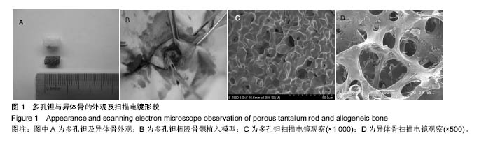
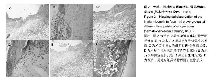
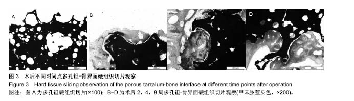
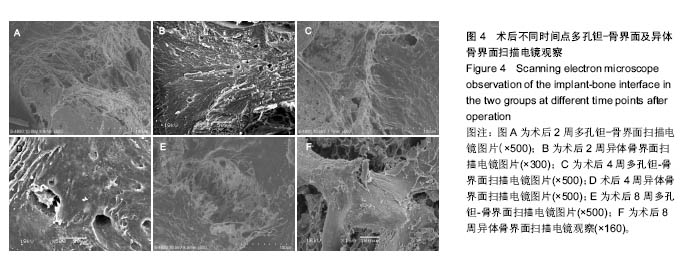
方法:实验采用自身对照方法,取日本大耳白兔制备双侧股骨髁骨缺损模型,同一动物左侧股骨内植入多孔钽棒为实验组,右侧股骨内植入异体骨为对照组。术后2,4,8 周于骨缺损部位取材,石蜡切片、硬组织切片光镜观察多孔钽与宿主骨交界处成骨形态特点;扫描电镜观察多孔钽-骨界面成骨特征;免疫组织化学检测整合素β1及纤维粘连蛋白的表达。
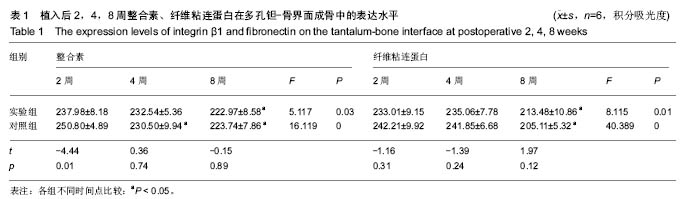
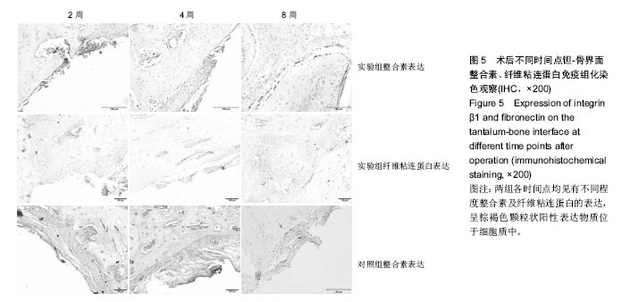
结果与结论:①多孔钽棒植入后与宿主骨结合紧密。石蜡切片苏木精-伊红染色结果显示:界面纤维膜早期疏松、较厚,晚期致密较薄,界面出现片状成骨;②硬组织切片观察:2周时钽-骨界面已出现新生骨及小血管并向孔隙内生长;4、8周时钽-宿主骨界面新生骨增多并与宿主骨连接成片,骨小梁成熟;③扫描电镜下可见成骨细胞在多孔钽表面及孔隙内生长,晚期骨质成熟并见板层骨;④免疫组化结果显示:多孔钽骨植入2周时实验组整合素 β1的表达显著低于对照组(P < 0.05);而纤维粘连蛋白的表达在两组之间比较差异均无显著性意义(P > 0.05);整合素及纤维粘连蛋白的表达在2,4,8周时均呈递减趋势;⑤结论:多孔钽有利于成骨细胞在其表面及孔隙内黏附,整合素β1及纤维粘连蛋白在成骨初期钽-骨界面表达增高,可能对早期成骨起促进作用,而在骨成熟期表达则降低,有利于骨整合及改建。

关键词: 生物材料, 骨生物材料, 多孔钽, 整合素, 纤维粘连蛋白, 钽-骨界面, 成骨

Abstract:

BACKGROUND: Previous studies have shown that home-made porous tantalum has non-toxicity and good biocompatibility, and can promote osteogenesis. Herein, we explore the mechanisms of tantalum-bone interface osseointegration.
OBJECTIVE: To observe the morphological characteristics and expressions of integrin β1 and fibronectin on the interface between porous tantalum and bone tissues after implantation into the right rabbit femur, and to evaluate the biological mechanisms of tantalum-bone interface osseointegration.
METHODS: Animal models of bilateral femoral condyle defects were made in Japanese big ear rabbits. Porous tantalum rod and allogeneic bone were respectively implanted into the left (experimental group) and right (control group) femur of rabbits. The animal specimens at the bone defect region were taken and made into paraffin sections and hard tissue sections at postoperative 2, 4, 8 weeks for morphological observation of new bone at the junction between the tantalum rod and host bone under light microscope, for osteogenic observation of the tantalum-bone interface under scanning electron microscope, and for immunohistochemical detection of integrin β1 and fibronectin expression.
RESULTS AND CONCLUSION: Porous tantalum was bonded closely with the host bone. The loose and thick fibrous capsule was observed in the early stage and became thinner in the late stage shown by hematoxylin-eosin staining. The new bone was visible on tantalum-bone interface. Hard tissue slicing observation showed that the new bone was seen on the porous tantalum-bone interface, blood capillaries grew into the pores at postoperative 2 weeks and the pores were full of new bone tissues at postoperative 4 and 8 weeks. Under the scanning electron microscope, the osteoblasts appeared on the tantalum surface and in the pores at the early stage, and bone maturation and lamellar bone were seen at the late stage. The immunohistochemical results showed that the expression of integrin β1 in the experimental group was significantly lower than that in the control group at postoperative 2 weeks (P < 0.05), but the expression of fibronectin had no significant difference between the two groups (P > 0.05). In addition, there was a decline trend in the expression of integrin β1 and fibronectin at postoperative 2, 4, 8 weeks. To conclude, the porous tantalum material is beneficial to enhance adhesion of osteoblasts on the surface and inside the micro-pores. Increased expression of integrin β1 and fibronectin on the tantalum-bone interface at early stage may promote early osteogenesis, while their decreased expression at bone maturing stage can promote osseointegration and bone remodeling.

Key words: Tantalum, Bone Transplantation, Biocompatible Materials, Tissue Engineering

中图分类号: