中国组织工程研究 ›› 2017, Vol. 21 ›› Issue (11): 1701-1706.doi: 10.3969/j.issn.2095-4344.2017.11.011

• 脊柱植入物 spinal implant • 上一篇    下一篇

下颈椎椎弓根钉进钉点与关节突间侧凹的三维定位关系

卢政好1,周菁华2,王卫国3   

  1. 南华大学附属南华医院,1脊柱外科,2放射科,湖南省衡阳市 421002;3中南大学湘雅三医院骨科,湖南省长沙市 410013
  • 修回日期:2017-01-06 出版日期:2017-04-18 发布日期:2017-05-06
  • 通讯作者: 王卫国,博士,主治医师,中南大学湘雅三医院骨科,湖南省长沙市 410013
  • 作者简介:卢政好,男,1975年生,辽宁省大石桥市人,汉族,2000年中南大学湘雅医学院毕业,硕士,副主任医师,主要从事脊柱外科方面的研究

Three-dimensional analysis of pedicle screw entry point and lateral concave between articular process in lower cervical vertebrae  

Lu Zheng-hao1, Zhou Jing-hua2, Wang Wei-guo3   

  1. 1Department of Spinal Surgery, Affiliated Nanhua Hospital, South China University, Hengyang 421002, Hunan Province, China; 2Department of Radiology, Affiliated Nanhua Hospital, South China University, Hengyang 421002, Hunan Province, China; 3Department of Orthopedics, the Third Xiangya Hospital, Central South University, Changsha 410013, Hunan Province, China
  • Revised:2017-01-06 Online:2017-04-18 Published:2017-05-06
  • Contact: Wang Wei-guo, M.D., Attending physician, Department of Orthopedics, the Third Xiangya Hospital, Central South University, Changsha 410013, Hunan Province, China
  • About author:Lu Zheng-hao, Master, Associate chief physician, Department of Spinal Surgery, Affiliated Nanhua Hospital, South China University, Hengyang 421002, Hunan Province, China

摘要:

文章快速阅读:


文题释义:
椎弓根钉进钉点:顾名思义就是经椎弓根固定时选择的螺钉入点,理想的椎弓根钉进钉点应位于椎弓根轴线在脊柱后柱的投影点上。正确选择进钉点是成功实行椎弓根钉固定技术的关键,因为它决定了进钉方向。目前,下胸椎和腰椎的椎弓根钉固定技术较为成熟。但在颈椎和中上胸椎,因为其椎弓根横径较为细小、周围毗邻关系复杂,所以导致该技术螺钉误置率较高,并发症较多,限制了临床应用。有鉴于此,进钉点的选择显得尤为重要。
关节突间侧凹:位于侧块外缘,为上下关节突间的凹陷部分,是下颈椎的固有结构。该解剖标志恒定、清晰可见、少有变异、未发现增生,而关节突增生后该结构更明显。下颈椎“关节突间侧凹”与椎弓根轴线在后柱的投影点有一定的定位关系。明确两者之间的定位关系,可为下颈椎椎弓根螺钉内固定提供一种安全、简便的进钉点选择方法。
 
摘要
背景:现有的下颈椎椎弓根钉固定技术均以关节突和侧块为参考标志,由于术者主观判断的差异,加之关节突增生的影响,进钉点的选择常有一定误差,易发生椎动脉或颈髓损伤等严重并发症,限制了该技术临床推广。目前临床上选择一个恒定的下颈椎椎弓根钉进钉点标志显得尤为重要。
目的:通过三维重建CT图像测量明确下颈椎椎弓根钉进钉点与“关节突间侧凹”的定位关系。
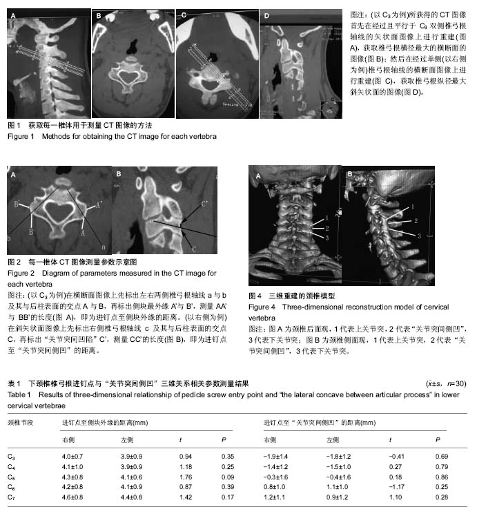
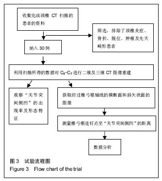
方法:对排除颈椎畸形的30例患者颈椎行三维重建CT扫描,观察“关节突间侧凹”的出现率和形态特征。在重建后的C3-C7特定CT图像上测定以下参数:①进钉点至侧块外缘的距离:经双侧椎弓根轴线横断面图像上椎弓根轴线在后柱表面投影点与侧块最外缘的距离;②进钉点至“关节突间侧凹”的距离:经单侧椎弓根轴线斜矢状面图像上椎弓根轴线在后柱表面投影点与“关节突间侧凹”的距离。统计每组参数的平均值和标准差,并比较各节段统计数据的差异。
结果与结论:①下颈椎“关节突间侧凹”明显,出现率为100%,解剖标志恒定且少有增生;②利用三维重建CT技术,可以成功获取C3-C7经双侧椎弓根轴线横断面和经单侧椎弓根轴线斜矢状面的图像;③在经双侧椎弓根轴线横断面图像上,C3-C7左右两侧进钉点至侧块外缘的距离分别为(4.1±0.9)mm和(4.3±0.9)mm,差异无显著性意义(P=0.609)。除了C3与C7、C4与C7,各节段同侧测量值的差异均无显著性意义(P > 0.05);④在经单侧椎弓根轴线斜矢状面的图像上,C3-C7左右两侧进钉点至“关节突间侧凹”的距离距离分别为(-0.3±1.7) mm和(-0.3±1.6) mm,差异无显著性意义(P=0.916)。除了C3与C4,各节段同侧测量值的差异均有显著性意义(P < 0.05);⑤以上结果提示,下颈椎椎弓根钉进钉点与“关节突间侧凹”在横断面上存在相对恒定的定位关系,而在矢状面上变异较大。
 
中国组织工程研究杂志出版内容重点:人工关节;骨植入物;脊柱骨折;内固定;数字化骨科;组织工程
ORCID: 0000-0001-9245-5587(卢政好)

关键词: 骨科植入物, 脊柱植入物, 下颈椎, 椎弓根螺钉, 内固定, 进钉点, 三维重建, CT, 测量, 关节突, 置钉准确性

Abstract:

BACKGROUND: The existing technique takes articular process and lateral mass as a reference mark. Due to differences of subjective judgment in operation and the impact of articular hyperplasia, some errors of screw entry point selection and serious complications such as vertebral artery or cervical spinal cord injury easily occurred, which limit the clinical application of the technique. At present, it is very important to select a constant reference mark of cervical pedicle screw entry point.

OBJECTIVE: Three-dimensional reconstruction CT images were made to measure the relationship between the lower cervical pedicle screw entry point and “the lateral concave between articular process”.
METHODS: Three-dimensional reconstruction of CT scan was performed in 30 patients with cervical deformity, and the occurrence rate and morphological characteristics of “the lateral concave between articular process” were observed. The following parameters were determined on specific reconstructed CT image of C3-C7: (1) the distance between pedicle axis projection point in the posterior surface and the outer edge of lateral mass in transversal section through bilateral pedicle axis and (2) the distance between the point and “the lateral concave between articular process” in oblique sagittal section through homolateral pedicle axis. Mean value and standard deviation were counted and statistics difference was compared.
RESULTS AND CONCLUSION: (1) “The lateral concave between articular process” in lower cervical vertebrae was a obvious and less constant proliferative anatomical landmark and its occurrence rate was 100%. (2) Using CT technology of 3D reconstruction, C3-C7 lower cervical transversal section through bilateral pedicle axis and oblique sagittal section through homolateral pedicle axis were successfully obtained. (3) In transversal section, the left and right distances between C3-C7 pedicle axis projection point in the posterior surface and the outer edge of lateral mass were (4.1±0.9) mm and (4.3±0.9) mm, and the difference was not statistically significant (P=0.609). Except for C3 and C7, C4 and C7, there was no significant difference in the measured values of the same side (P > 0.05). (4) In oblique sagittal section, the left and right distances between C3-C7 point and “the lateral concave between articular process” were (-0.3±1.7) mm and (-0.3±1.6) mm, and the difference was not statistically significant (P=0.916). Except for C3 and C4, there was significant difference in the measured values of the same side (P < 0.05). (5) The above results suggest that the lower cervical pedicle entry point and “the lateral concave between articular process” have a relatively constant orientation relationship in transversal section and a large variation in the sagittal section.

中国组织工程研究杂志出版内容重点:人工关节;骨植入物;脊柱骨折;内固定;数字化骨科;组织工程

Key words: Cervical Vertebrae, Bone Nails, Internal Fixators, Tissue Engineering

中图分类号: