| [1] 戚大春,安新荣.全髋关节置换假体位置的确定方法及生物力学特性[J].中国组织工程研究,2016,20(26):3811-3816. [2] Clavé A, Fazilleau F, Dumser D, et al. Efficacy of tranexamic acid on blood loss after primary cementless total hip replacement with rivaroxaban thromboprophylaxis: A case-control study in 70 patients. Orthop Traumatol Surg Res. 2012;98(5):484-490. [3] Alshryda S, Mason J, Vaghela M, et al. Topical (intra-articular) tranexamic acid reduces blood loss and transfusion rates following total knee replacement: a randomized controlled trial (TRANX-K). J Bone Joint Surg Am. 2013;95(21):1961-1968. [4] 张波,庞清江,章海均,等.全膝关节置换术后隐性失血的研究进展[J].中国骨伤,2012,25(9):788-792. [5] [5] Alshryda S, Mason J, Sarda P, et al. Topical (intra-articular) tranexamic acid reduces blood loss and transfusion rates following total hip replacement: a randomized controlled trial (TRANX-H). J Bone Joint Surg Am. 2013;95(21):1969-1974. [6] Irwin A, Khan SK, Jameson SS, et al. Oral versus intravenous tranexamic acid in enhanced-recovery primary total hip and knee replacement: results of 3000 procedures. Bone Joint J. 2013;95-B(11):1556-1561. [7] 谢锦伟,岳辰,裴福兴.氨甲环酸在全髋关节置换术中的有效性与安全性研究进展[J].中国矫形外科杂志, 2014,22(20):1856-1860.[8] Bao N, Zhou L, Cong Y, et al. Free fatty acids are responsible for the hidden blood loss in total hip and knee arthroplasty. Med Hypotheses. 2013;81(1):104-107. [9] Li J, Zhou Y, Jing J, et al. Comparison of effects of two anticoagulants on hidden blood loss after total hip arthroplasty. Zhongguo Xiu Fu Chong Jian Wai Ke Za Zhi. 2013;27(4): 432-435.[10] 李宝山,冷灵,李成.髓外和髓内固定及髋关节置换治疗高龄股骨粗隆间骨折隐性失血的特点分析[J].实用医学杂志, 2015(18): 3004-3007[11] 庞向华,欧阳建江,欧兆强,等.隐性失血与全髋关节置换术后髋关节功能的相关性及影响因素[J].广东医学,2013,34(3): 420-422. [12] 米尔阿里木•木尔提扎,赵巍,王利,等.患者体重指数对全髋关节置换手术时间的影响研究[J].实用骨科杂志,2014, 20(10): 938-941. [13] 王世海,颜冰.肥胖对全髋关节置换术后隐性失血的影响[J].深圳中西医结合杂志,2014,24(9):119-120. [14] Kelly EG, Cashman JP, Imran FH, et al. Systematic review and meta-analysis of closed suction drainage versus non-drainage in primary hip arthroplasty. Surg Technol Int. 2014;24:295-301.[15] 李军,荆珏华,史占军,等.利伐沙班对全髋关节置换术隐性出血影响的病例对照研究[J].中国骨伤,2014,27(1):34-37. [16] 张晓强,高菲菲,王战朝,等.膝伤活血灵口服配合低分子肝素钙皮下注射对全膝关节置换术后隐性失血的影响[J].中医正骨, 2014, 26(4):23-25.[17] 连奎,戚超,于腾,等.骨水泥与非骨水泥型人工全髋关节置换治疗肾移植术后股骨头缺血性坏死的近期疗效比较[J].中国修复重建外科杂志,2013,27(12):1409-1413.[18] 许超,孙鹏飞,郭永园,等.一期翻修并植骨治疗THR术后感染伴髋臼骨缺损30例临床分析[J].中国实用外科杂志, 2014,34(S1): 47-48.[19] 黄文泽,彭昊,方洪松,等.大头陶瓷-陶瓷人工全髋关节置换术治疗成人先天性髋关节发育不良[J].临床骨科杂志, 2013,16(2): 151-154.[20] 袁宏,陆琳松,钟惠琴,等.唑来膦酸对骨质疏松患者全髋关节置换术后假体周围骨密度的影响[J].中华关节外科杂志(电子版), 2014,1(3):278-285.[21] 周定,臧谋圣,胡勇,等.骨水泥与非骨水泥股骨柄在初次全髋关节置换术后疗效的Meta分析[J].中华关节外科杂志(电子版), 2014,8(5):635-642.[22] 屈瑾,雷新玮,展影,等.髋关节表面置换术假体柄固定方式对股骨近端骨密度及应力影响[J].中华医学杂志, 2014,94(35): 2769-2771.[23] 李宁远,龚亚莉,刘煊文,等.不同材料人工髋关节假体对骨界面应力分布及生物力学的影响[J].中国组织工程研究, 2016,20(9): 1268-1274. [24] Pinheiro AL, Santos NR, Oliveira PC, et al. The efficacy of the use of IR laser phototherapy associated to biphasic ceramic graft and guided bone regeneration on surgical fractures treated with wire osteosynthesis: a comparative laser fluorescence and Raman spectral study on rabbits. Lasers Med Sci. 201;28(3):815-822. [25] Pinheiro AL, Santos NR, Oliveira PC, et al. The efficacy of the use of IR laser phototherapy associated to biphasic ceramic graft and guided bone regeneration on surgical fractures treated with miniplates: a Raman spectral study on rabbits. Lasers Med Sci. 2013;28(2):513-518. [26] Pinheiro AL, Aciole GT, Ramos TA, et al. The efficacy of the use of IR laser phototherapy associated to biphasic ceramic graft and guided bone regeneration on surgical fractures treated with miniplates: a histological and histomorphometric study on rabbits. Lasers Med Sci. 2014;29(1):279-288. [27] Lopes CB, Pacheco MT, Silveira L Jr, et al. The effect of the association of NIR laser therapy BMPs, and guided bone regeneration on tibial fractures treated with wire osteosynthesis: Raman spectroscopy study. J Photochem Photobiol B. 2007;89(2-3):125-130. [28] Lopes CB, Pacheco MT, Silveira L Jr, et al. The effect of the association of near infrared laser therapy, bone morphogenetic proteins, and guided bone regeneration on tibial fractures treated with internal rigid fixation: a Raman spectroscopic study. J Biomed Mater Res A. 2010;94(4): 1257-1263.[29] Schneider D, Weber FE, Grunder U, et al. A randomized controlled clinical multicenter trial comparing the clinical and histological performance of a new, modified polylactide-co-glycolide acid membrane to an expanded polytetrafluorethylene membrane in guided bone regeneration procedures. Clin Oral Implants Res. 2014;25(2):150-158. [30] Annen BM, Ramel CF, Hämmerle CH, et al. Use of a new cross-linked collagen membrane for the treatment of peri-implant dehiscence defects: a randomised controlled double-blinded clinical trial. Eur J Oral Implantol. 2011;4(2): 87-100. [31] Carpio L, Loza J, Lynch S, et al. Guided bone regeneration around endosseous implants with anorganic bovine bone mineral. A randomized controlled trial comparing bioabsorbable versus non-resorbable barriers. J Periodontol. 2000;71(11):1743-1749. [32] Ronda M, Rebaudi A, Torelli L, et al. Expanded vs. dense polytetrafluoroethylene membranes in vertical ridge augmentation around dental implants: a prospective randomized controlled clinical trial. Clin Oral Implants Res. 2014;25(7):859-866. [33] Hämmerle CH, Lang NP. Single stage surgery combining transmucosal implant placement with guided bone regeneration and bioresorbable materials. Clin Oral Implants Res. 2001;12(1):9-18. [34] Todisco M. Early loading of implants in vertically augmented bone with non-resorbable membranes and deproteinised anorganic bovine bone. An uncontrolled prospective cohort study. Eur J Oral Implantol. 2010;3(1):47-58. [35] Chiapasco M, Zaniboni M. Clinical outcomes of GBR procedures to correct peri-implant dehiscences and fenestrations: a systematic review. Clin Oral Implants Res. 2009;20 Suppl 4:113-123.[36] 刘文广,刘胜厚,殷庆丰,等.水泥型人工髋关节置换治疗老年EvansⅠ-Ⅲ型粗隆间骨折股骨假体的生物力学特性[J].中国医学科学院学报,2013,35(1):108-111.[37] 谢锦伟,岳辰,裴福兴.氨甲环酸在全髋关节置换术中的有效性与安全性研究进展[J].中国矫形外科杂志,2014, 22(20):1856- 1860.[38] 张晓强,高菲菲,王战朝,等.膝伤活血灵口服配合低分子肝素钙皮下注射对全膝关节置换术后隐性失血的影响[J].中医正骨, 2014, 26(4):23-25. [39] Li J, Zhou Y, Jing J, et al. Comparison of effects of two anticoagulants on hidden blood loss after total hip arthroplasty. Zhongguo Xiu Fu Chong Jian Wai Ke Za Zhi. 2013;27(4): 432-435.[40] 袁磊,包倪荣,赵建宁.髋关节置换术后隐性失血的新进展[J].中国骨伤,2015,28(4):378-382. |
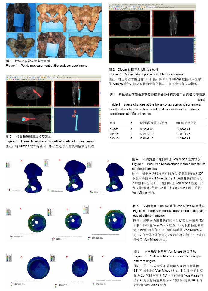
.jpg)


.jpg)