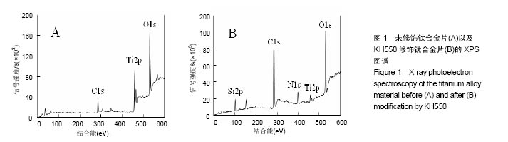
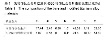
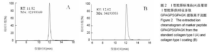
| [1]de Andrade DP, de Vasconcellos LM, Carvalho IC, et al. Titanium-35niobium alloy as a potential material for biomedical implants: In vitro study. Mater Sci Eng C Mater Biol Appl. 2015;56:538-544.[2]Kolmas J, Oledzka E, Sobczak M, et al. Nanocrystalline hydroxyapatite doped with selenium oxyanions: a new material for potential biomedical applications.Mater Sci Eng C Mater Biol Appl. 2014;39:134-142.[3]Joo JY, Amin ML, Rajangam T, et al. Fibrinogen as a promising material for various biomedical applications. Molecular & Cellular Toxicology. 2015; 11(1):1-9.[4]Mosadegh B, Xiong G, Dunham S, et al. Current progress in 3D printing for cardiovascular tissue engineering. Biomed Mater. 2015;10(3):034002.[5]Farnoush H, Aghazadeh Mohandesi J, Çimeno?lu H. Micro-scratch and corrosion behavior of functionally graded HA-TiO2 nanostructured composite coatings fabricated by electrophoretic deposition. J Mech Behav Biomed Mater. 2015;46:31-40.[6]Himics L, Tóth S, Veres M, et al. Effective implantation of light emitting centers by plasma immersion ion implantation and focused ion beam methods into nanosized diamond. Applied Surface Science. 2015; 328:577-582.[7]Quan Z, Ni E, Ogasawara Y, et al. Nano-size multiple metal oxide anode electrodes synthesized from layered double hydroxides - Electrochemical reaction mechanism and surface morphology change during reaction with lithium ion. Solid State Ionics. 2014; 268: 268-272. [8]Liang CY, Zhong X, Wang HS, et al. Femtosecond laser induced micropatterns and in-situ deposition of Ca/P phase and collagen on Ti surface. Materials Chemistry and Physics. 2015; 158: 115-120.[9]Hauser J, Koeller M, Bensch S, et al. Plasma mediated collagen-I-coating of metal implant materials to improve biocompatibility. J Biomed Mater Res A. 2010;94(1):19-26.[10]Schulz MC, Korn P, Stadlinger B, et al. Coating with artificial matrices from collagen and sulfated hyaluronan influences the osseointegration of dental implants.J Mater Sci Mater Med. 2014;25(1):247-258.[11]Ricard-Blum S, Ruggiero F. The collagen superfamily: from the extracellular matrix to the cell membrane. Pathol Biol (Paris). 2005;53(7):430-442.[12]Huang CY, Kuo JM, Wu SJ, et al. Isolation and characterization of fish scale collagen from tilapia (Oreochromis sp.) by a novel extrusion-hydro-extraction process. Food Chem. 2016;190:997-1006.[13]Martínezortiz MA, Hernándezfuentes AD, Pimentelgonzález DJ, et al. Extraction and characterization of collagen from rabbit skin: partial characterization. CyTA - Journal of Food. 2015; 13(2):253-258.[14]Shanmugam G, Reddy SMM, Madhan B,et al. Method of addition of acetonitrile influences the structure and stability of collagen. Process Biochemistry. 2014;49(2): 210-216.[15]Li GY, Fukunaga S, Takenouchi K, et al. Comparative study of the physiological properties of collagen, gelatin and collagen hydrolysate as cosmetic materials. Int J Cosmet Sci. 2005; 27(2):101-106.[16]Hauser J, Ring A, Schaffran A, et al. In vivo analysis of tissue response to plasma-treated collagen-I-coated titanium alloys. Eur Surg Res. 2009;43(3):262-268.[17]Felgueiras HP, Sommerfeld SD, Murthy NS, et al. Poly(NaSS) functionalization modulates the conformation of fibronectin and collagen type I to enhance osteoblastic cell attachment onto Ti6Al4V. Langmuir. 2014;30(31):9477-9483.[18]Gigante A, Bevilacqua C, Cappella M, et al. Engineered articular cartilage: influence of the scaffold on cell phenotype and proliferation. J Mater Sci Mater Med. 2003;14(8): 713-716.[19]Buma P, Pieper JS, van Tienen T, et al. Cross-linked type I and type II collagenous matrices for the repair of full-thickness articular cartilage defects--a study in rabbits. Biomaterials. 2003;24(19):3255-3263.[20]Nehrer S, Breinan HA, Ramappa A, et al. Matrix collagen type and pore size influence behaviour of seeded canine chondrocytes. Biomaterials. 1997;18(11):769-776.[21]Su PJ, Chen WL, Li TH, et al. The discrimination of type I and type II collagen and the label-free imaging of engineered cartilage tissue. Biomaterials. 2010;31(36):9415-9421.[22]Chang KY, Hung LH, Chu IM, et al. The application of type II collagen and chondroitin sulfate grafted PCL porous scaffold in cartilage tissue engineering. J Biomed Mater Res A. 2010; 92(2):712-723. [23]Cao YL, Liu T, Pang J, et al. Glucan HBP-A increase type II collagen expression of chondrocytes in vitro and tissue engineered cartilage in vivo. Chin J Integr Med. 2015;21(3): 196-203.[24]Kontturi LS, Järvinen E, Muhonen V, et al. An injectable, in situ forming type II collagen/hyaluronic acid hydrogel vehicle for chondrocyte delivery in cartilage tissue engineering. Drug Deliv Transl Res. 2014;4(2):149-158.[25]Yao Y, Ma YZ, Qin M, et al. NHS-ester functionalized poly(PEGMA) brushes on silicon surface for covalent protein immobilization. Colloids Surf B Biointerfaces. 2008;66(2): 233-239.[26]孙爱梅,张贵锋,倪文,等. 胶原蛋白降解物高效液相色谱/质谱联用分析[J]. 中国生物工程杂志, 2005, 25(2): 66-72.[27]张贵锋,刘涛,王前,等. 高效液相色谱/质谱法识别不同明胶酶解产物中特征多肽[J]. 分析化学, 2008, 36(11): 1499-1504.[28]Webster TJ, Ejiofor JU. Increased osteoblast adhesion on nanophase metals: Ti, Ti6Al4V, and CoCrMo. Biomaterials. 2004;25(19):4731-4739.[29]Shah FA, Trobos M, Thomsen P, et al. Commercially pure titanium (cp-Ti) versus titanium alloy (Ti6Al4V) materials as bone anchored implants - Is one truly better than the other. Mater Sci Eng C Mater Biol Appl. 2016;62:960-966.[30]Crespo L, Hierro-Oliva M, Barriuso S, et al. On the interactions of human bone cells with Ti6Al4V thermally oxidized by means of laser shock processing. Biomed Mater. 2016;11(1):015009.[31]Calzado-Martín A, Crespo L, Saldaña L,et al. Human bone-lineage cell responses to anisotropic Ti6Al4V surfaces are dependent on their maturation state. J Biomed Mater Res A. 2014;102(9):3154-3166.[32]Barão VA, Mathew MT, Assunção WG, et al. Stability of cp-Ti and Ti-6Al-4V alloy for dental implants as a function of saliva pH - an electrochemical study. Clin Oral Implants Res. 2012; 23(9):1055-1062.[33]Mohseni E, Zalnezhad E, Bushroa AR.Comparative investigation on the adhesion of hydroxyapatite coating on Ti–6Al–4V implant: A review paper. International Journal of Adhesion & Adhesives. 2014;48(1):238-257.[34]Durdu S, Deniz OF, Kutbay I, et al.Characterization and formation of hydroxyapatite on Ti6Al4V coated by plasma electrolytic oxidation. Journal of Alloys and Compounds. 2013; 551: 422-429.[35]Arifin A, Sulong AB, Muhamad N, et al. Material processing of hydroxyapatite and titanium alloy (HA/Ti) composite as implant materials using powder metallurgy: a review. Materials & Design. 2014; 55(6): 165-175. [36]Benea L, Mardare-Danaila E, Mardare M, et al. Preparation of titanium oxide and hydroxyapatite on Ti–6Al–4V alloy surface and electrochemical behaviour in bio-simulated fluid solution. Corrosion Science. 2014; 80(3): 331-338.[37]Liu Y, Cui H, Zhuang X, et al. Nano-hydroxyapatite surfaces grafted with electroactive aniline tetramers for bone-tissue engineering. Macromol Biosci. 2013;13(3):356-365.[38]Raghavan RN, Muthukumar T, Somanathan N, et al. Biomimetic mineralization of novel silane crosslinked collagen. Mater Sci Eng C Mater Biol Appl. 2013;33(4):1983-1988.[39]Zhu H, Hu C, Zhang F, et al. Preparation and antibacterial property of silver-containing mesoporous 58S bioactive glass. Mater Sci Eng C Mater Biol Appl. 2014;42:22-30.[40]Chiu CK, Ferreira J, Luo TJ, et al. Direct scaffolding of biomimetic hydroxyapatite-gelatin nanocomposites using aminosilane cross-linker for bone regeneration. J Mater Sci Mater Med. 2012;23(9):2115-2126.[41]张杭州, 孙羽, 王琳, 等. 羟基磷灰石/TiO2纳米管复合物的生物相容性[J]. 中国组织工程研究, 2014, 18(3): 335-340.[42]鲁雄, 冯波, 翁杰, 等. 生物材料表面微纳结构对成骨相关细胞的影响[J]. 中国材料进展, 2013, 32(10): 612-622.[43]Higuchi A, Ling QD, Chang Y, et al. Physical cues of biomaterials guide stem cell differentiation fate. Chem Rev. 2013;113(5):3297-3328.[44]Lee TT, García JR, Paez JI, et al. Light-triggered in vivo activation of adhesive peptides regulates cell adhesion, inflammation and vascularization of biomaterials. Nat Mater. 2015;14(3):352-360.[45]Shafiq M, Jung Y, Kim SH. Insight on stem cell preconditioning and instructive biomaterials to enhance cell adhesion, retention, and engraftment for tissue repair. Biomaterials. 2016;90:85-115. [46]Spicer V, Ezzati P, Neustaeter H, et al. 3D HPLC-MS with Reversed-Phase Separation Functionality in All Three Dimensions for Large-Scale Bottom-Up Proteomics and Peptide Retention Data Collection. Anal Chem. 2016;88(5):2847-5285.[47]Tscheliessnig AL, Konrath J, Bates R, et al. Host cell protein analysis in therapeutic protein bioprocessing - methods and applications. Biotechnol J. 2013;8(6):655-670.[48]Hou J, Tobe BT, Lo F, et al. Combined total proteomic and phosphoproteomic analysis of human pluripotent stem cells. Methods Mol Biol. 2013;1029:163-189.[49]Köcher T, Pichler P, Swart R, et al. Analysis of protein mixtures from whole-cell extracts by single-run nanoLC-MS/MS using ultralong gradients. Nat Protoc. 2012;7(5):882-890. |
.jpg)


.jpg)