中国组织工程研究 ›› 2017, Vol. 21 ›› Issue (8): 1172-1177.doi: 10.3969/j.issn.2095-4344.2017.08.005
• 口腔组织构建 oral tissue construction • 上一篇 下一篇
富自体浓缩生长因子膜在兔下颌骨牵张成骨中的作用
田璐鸣1,辛增玺1,马云胜2
- 1辽宁医学院附属第二医院口腔外科,辽宁省锦州市 121001;2锦州医科大学发育生物学教研室,辽宁省锦州市 121000
Effect of concentrated growth factor fibrin membrane during distraction osteogenesis of the rabbit mandible
Tian Lu-ming1, Xin Zeng-xi1, Ma Yun-sheng2
- 1Department of Oral Surgery, the Second Affiliated Hospital of Liaoning Medical University, Jinzhou 121001, Liaoning Province, China; 2Department of Developmental Biology, Jinzhou Medical University, Jinzhou 121000, Liaoning Province, China
摘要:
文章快速阅读:
.jpg) 文题释义:
牵张成骨:是一种内源性骨组织工程技术,是通过将骨骼切开,在切骨线两侧安放特制的牵张器,经过一定的延迟期后(5-7 d),缓慢牵张切骨间隙(1-1.5 mm/d),使切骨间隙不断增宽,并激发机体组织再生的潜力,在牵张间隙内不断形成新生骨组织,同时使骨骼周围的肌肉、神经、血管、皮肤等同期延长,从而达到延长骨骼的目的。
富自体浓缩生长因子纤维蛋白凝胶(液体):是一种新型血液提取物,是继富血小板血浆和富血小板纤维蛋白之后的第3代血浆提取物;浓缩生长因子在细胞迁移,细胞增殖和血管生成组织再生方面有至关重要的作用,如富血小板血浆,富血小板纤维蛋白,富自体浓缩生长因子已被用于重建骨缺损。
文题释义:
牵张成骨:是一种内源性骨组织工程技术,是通过将骨骼切开,在切骨线两侧安放特制的牵张器,经过一定的延迟期后(5-7 d),缓慢牵张切骨间隙(1-1.5 mm/d),使切骨间隙不断增宽,并激发机体组织再生的潜力,在牵张间隙内不断形成新生骨组织,同时使骨骼周围的肌肉、神经、血管、皮肤等同期延长,从而达到延长骨骼的目的。
富自体浓缩生长因子纤维蛋白凝胶(液体):是一种新型血液提取物,是继富血小板血浆和富血小板纤维蛋白之后的第3代血浆提取物;浓缩生长因子在细胞迁移,细胞增殖和血管生成组织再生方面有至关重要的作用,如富血小板血浆,富血小板纤维蛋白,富自体浓缩生长因子已被用于重建骨缺损。
文题释义:
牵张成骨:是一种内源性骨组织工程技术,是通过将骨骼切开,在切骨线两侧安放特制的牵张器,经过一定的延迟期后(5-7 d),缓慢牵张切骨间隙(1-1.5 mm/d),使切骨间隙不断增宽,并激发机体组织再生的潜力,在牵张间隙内不断形成新生骨组织,同时使骨骼周围的肌肉、神经、血管、皮肤等同期延长,从而达到延长骨骼的目的。
富自体浓缩生长因子纤维蛋白凝胶(液体):是一种新型血液提取物,是继富血小板血浆和富血小板纤维蛋白之后的第3代血浆提取物;浓缩生长因子在细胞迁移,细胞增殖和血管生成组织再生方面有至关重要的作用,如富血小板血浆,富血小板纤维蛋白,富自体浓缩生长因子已被用于重建骨缺损。摘要
背景:富自体浓缩生长因子膜可参与调节代谢,已被用于骨缺损的重建。研究发现在牵张成骨的新骨缝附近的成骨细胞、间质细胞及骨细胞的胞质中均有骨保护素和核因子ΚB受体活化因子配体表达。
目的:分析骨矫形作用的机制及富自体浓缩生长因子膜对兔下颌骨牵张成骨的促进意义。
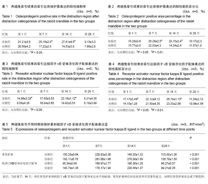
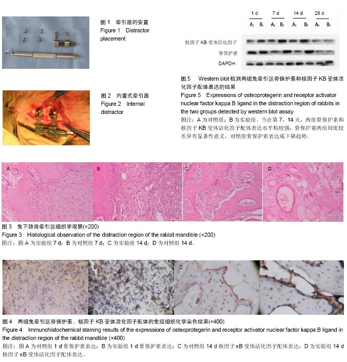
方法:随机选取24只大白兔建立兔下颌骨牵张成骨模型;对照组行左单侧下颌骨牵张成骨;实验组将富自体浓缩生长因子膜固定于牵张成骨器内侧面并且完全包绕牵张间隙,再行右单侧下颌骨牵张成骨;共延长 6 mm。在固定期第1,7,14及28天分别处死动物,获取双侧下颌骨行组织学苏木精-伊红染色、免疫印迹法Western blot法和免疫组织化学检测核因子ΚB受体活化因子配体及骨保护素在新生骨中的表达情况;观察和对比两组间牵张间隙内成骨效果。
结果与结论:牵引后第1,7,14天骨保护素表达的阳性细胞率和阳性面积百分比实验组均显著高于对照组(P < 0.05或P < 0.01);牵引后第1,14天核因子ΚB受体活化因子配体表达的阳性细胞率和阳性面积百分比实验组均显著高于对照组(P < 0.05或P < 0.01);Western blot法检测核因子ΚB受体活化因子配体/骨保护素比值对照组较实验组要高(P < 0.01)。结果说明,富自体浓缩生长因子膜可促进兔下颌骨牵张成骨间隙内的新骨形成和矿化,说明富自体浓缩生长因子膜是促进下颌骨牵张成骨的有效手段。
中国组织工程研究杂志出版内容重点:组织构建;骨细胞;软骨细胞;细胞培养;成纤维细胞;血管内皮细胞;骨质疏松;组织工程
ORCID: 0000-0003-1583-7893(田璐鸣)
中图分类号:
.jpg) 文题释义:
牵张成骨:是一种内源性骨组织工程技术,是通过将骨骼切开,在切骨线两侧安放特制的牵张器,经过一定的延迟期后(5-7 d),缓慢牵张切骨间隙(1-1.5 mm/d),使切骨间隙不断增宽,并激发机体组织再生的潜力,在牵张间隙内不断形成新生骨组织,同时使骨骼周围的肌肉、神经、血管、皮肤等同期延长,从而达到延长骨骼的目的。
富自体浓缩生长因子纤维蛋白凝胶(液体):是一种新型血液提取物,是继富血小板血浆和富血小板纤维蛋白之后的第3代血浆提取物;浓缩生长因子在细胞迁移,细胞增殖和血管生成组织再生方面有至关重要的作用,如富血小板血浆,富血小板纤维蛋白,富自体浓缩生长因子已被用于重建骨缺损。
文题释义:
牵张成骨:是一种内源性骨组织工程技术,是通过将骨骼切开,在切骨线两侧安放特制的牵张器,经过一定的延迟期后(5-7 d),缓慢牵张切骨间隙(1-1.5 mm/d),使切骨间隙不断增宽,并激发机体组织再生的潜力,在牵张间隙内不断形成新生骨组织,同时使骨骼周围的肌肉、神经、血管、皮肤等同期延长,从而达到延长骨骼的目的。
富自体浓缩生长因子纤维蛋白凝胶(液体):是一种新型血液提取物,是继富血小板血浆和富血小板纤维蛋白之后的第3代血浆提取物;浓缩生长因子在细胞迁移,细胞增殖和血管生成组织再生方面有至关重要的作用,如富血小板血浆,富血小板纤维蛋白,富自体浓缩生长因子已被用于重建骨缺损。