[1] 杨思敏,王新卫.自体骨移植修复骨缺损的临床研究进展[J].中国疗养医学,2019,28(9):945-948.
[2] REZAEI M, FARHADIAN M, RASHIDI AM, et al. Nano-Biphasic Calcium Phosphate Ceramic for the Repair of Bone Defects. J Craniofac Surg. 2018; 29(6):e543-e548.
[3] GARRIDO CA, LOBO SE, TURÍBIO FM, et al. Biphasic calcium phosphate bioceramics for orthopaedic reconstructions: clinical outcomes. Int J Biomater. 2011;2011:129727.
[4] MANGANO FG, ZECCA PA, VAN NOORT R, et al. Custom-Made Computer-Aided-Design/Computer-Aided-Manufacturing Biphasic Calcium-Phosphate Scaffold for Augmentation of an Atrophic Mandibular Anterior Ridge. Case Rep Dent. 2015;2015:941265.
[5] ZHANG Z, WANG P, LI X, et al. Reconstruction of mandibular bone defects using biphasic calcium phosphate bone substitutes with simultaneous implant placement in mini-swine: A pilot in vivo study. J Biomed Mater Res B Appl Biomater. 2019;107(6):2071-2079.
[6] BACAKOVA L, ZARUBOVA J, TRAVNICKOVA M, et al. Stem cells: their source, potency and use in regenerative therapies with focus on adipose-derived stem cells - a review. Biotechnol Adv. 2018;36(4):1111-1126.
[7] AL-GHADBAN S, BUNNELL BA. Adipose Tissue-Derived Stem Cells: Immunomodulatory Effects and Therapeutic Potential. Physiology (Bethesda). 2020;35(2):125-133.
[8] WAGNER JM, REINKEMEIER F, WALLNER C, et al. Adipose-Derived Stromal Cells Are Capable of Restoring Bone Regeneration After Post-Traumatic Osteomyelitis and Modulate B-Cell Response. Stem Cells Transl Med. 2019; 8(10):1084-1091.
[9] MAO SH, CHEN CH, CHEN CT. Osteogenic potential of induced pluripotent stem cells from human adipose-derived stem cells. Stem Cell Res Ther. 2019;10(1):303.
[10] DUFRANE D, DOCQUIER PL, DELLOYE C, et al. Scaffold-free Three-dimensional Graft From Autologous Adipose-derived Stem Cells for Large Bone Defect Reconstruction: Clinical Proof of Concept. Medicine (Baltimore). 2015;94(50):e2220.
[11] PADUANO F, MARRELLI M, AMANTEA M, et al. Adipose Tissue as a Strategic Source of Mesenchymal Stem Cells in Bone Regeneration: A Topical Review on the Most Promising Craniomaxillofacial Applications. Int J Mol Sci. 2017; 18(10):2140.
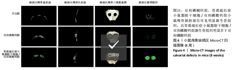
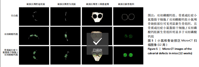
[12] 彭双麟,姚志浩,罗道文,等.多孔双相磷酸钙陶瓷修复骨质疏松症大鼠颅骨极量缺损的实验研究[J].口腔医学研究,2019,35(4):377-381.
[13] SANTOS PS, CESTARI TM, PAULIN JB, et al. Osteoinductive porous biphasic calcium phosphate ceramic as an alternative to autogenous bone grafting in the treatment of mandibular bone critical-size defects. J Biomed Mater Res B Appl Biomater. 2018;106(4):1546-1557.
[14] SHEU SY, HSU YK, CHUANG MH, et al. Enhanced Bone Formation in Osteoporotic Mice by a Novel Transplant Combined with Adipose-derived Stem Cells and Platelet-rich Fibrin Releasates. Cell Transplant. 2020;29: 963689720927398.
[15] 唐宇星,赵庆,杨中萌,等.聚乳酸共聚物复合脂肪干细胞对骨质疏松性骨折愈后生物力学的影响[J].中国组织工程研究,2017,21(10):1577-1582.
[16] WANG L, HUANG C, LI Q, et al. Osteogenic differentiation potential of adipose-derived stem cells from ovariectomized mice. Cell Prolif. 2017;50(2): e12328.
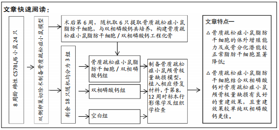
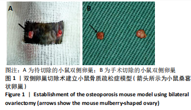
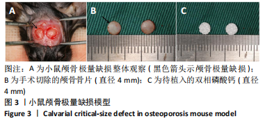
[17] 罗道文,黄馗,王雷,等.C57小鼠骨质疏松症伴颅骨极量缺损模型的建立[J].中国组织工程研究,2017,21(36):5793-5798.
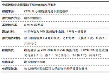
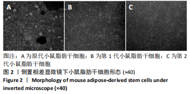
[18] 黄成龙,黎庆,王雷,等.两种方法培养骨质疏松症小鼠脂肪干细胞的比较[J].中国组织工程研究,2017,21(29):4654-4659.
[19] WUBNEH A, TSEKOURA EK, AYRANCI C, et al. Current state of fabrication technologies and materials for bone tissue engineering. Acta Biomater. 2018;80:1-30.
[20] ATASOY-ZEYBEK A, KOSE GT. Gene Therapy Strategies in Bone Tissue Engineering and Current Clinical Applications. Adv Exp Med Biol. 2018; 1119:85-101.
[21] AHMED S, CHAUHAN VM, GHAEMMAGHAMI AM, et al. New generation of bioreactors that advance extracellular matrix modelling and tissue engineering. Biotechnol Lett. 2019;41(1):1-25.
[22] GRIMM D, EGLI M, KRÜGER M, et al. Tissue Engineering Under Microgravity Conditions-Use of Stem Cells and Specialized Cells. Stem Cells Dev. 2018; 27(12):787-804.
[23] CHANDRA P, ATALA A. Engineering blood vessels and vascularized tissues: technology trends and potential clinical applications. Clin Sci (Lond). 2019; 133(9):1115-1135.
[24] ZUK PA, ZHU M, MIZUNO H, et al. Multilineage cells from human adipose tissue: implications for cell-based therapies. Tissue Eng. 2001;7(2):211-228.
[25] VAN ESTERIK FA, ZANDIEH-DOULABI B, KLEVERLAAN CJ, et al. Enhanced Osteogenic and Vasculogenic Differentiation Potential of Human Adipose Stem Cells on Biphasic Calcium Phosphate Scaffolds in Fibrin Gels. Stem Cells Int. 2016;2016:1934270.
[26] LIM HC, ZHANG ML, LEE JS, et al. Effect of different hydroxyapatite:β-tricalcium phosphate ratios on the osteoconductivity of biphasic calcium phosphate in the rabbit sinus model. Int J Oral Maxillofac Implants. 2015; 30(1):65-72.
[27] PRIPATNANONT P, PRASERTTHAM P, SUTTAPREYASRI S, et al. Bone Regeneration Potential of Biphasic Nanocalcium Phosphate with High Hydroxyapatite/Tricalcium Phosphate Ratios in Rabbit Calvarial Defects. Int J Oral Maxillofac Implants. 2016;31(2):294-303.
[28] YANG C, UNURSAIKHAN O, LEE JS, et al. Osteoconductivity and biodegradation of synthetic bone substitutes with different tricalcium phosphate contents in rabbits. J Biomed Mater Res B Appl Biomater. 2014;102(1):80-88.
[29] GU BK, CHOI DJ, PARK SJ, et al. 3D Bioprinting Technologies for Tissue Engineering Applications. Adv Exp Med Biol. 2018;1078:15-28.
[30] CANCIANI E, DELLAVIA C, FERREIRA LM, et al. Human Adipose-Derived Stem Cells on Rapid Prototyped Three-Dimensional Hydroxyapatite/Beta-Tricalcium Phosphate Scaffold. J Craniofac Surg. 2016;27(3):727-732. |