中国组织工程研究 ›› 2017, Vol. 21 ›› Issue (4): 527-531.doi: 10.3969/j.issn.2095-4344.2017.04.006

• 软骨组织构建 cartilage tissue construction • 上一篇    下一篇

肿瘤坏死因子α诱导大鼠软骨细胞凋亡模型的建立及鉴定

陈后煌1,邵  翔1,李  俐2,吴明霞2,李西海1   

  1. 1福建中医药大学中西医结合研究院,福建省福州市  350122;2福建中医药大学附属第二人民医院,福建省福州市  350003
  • 收稿日期:2016-12-06 出版日期:2017-02-08 发布日期:2017-03-13
  • 通讯作者: 通讯作者:吴明霞,博士,主任医师,教授,福建中医药大学附属第二人民医院,福建省福州市 350003 并列通讯作者:李西海,博士,副教授,福建中医药大学中西医结合研究院,福建省福州市 350122
  • 作者简介:陈后煌,男,1990年生,江西省丰城市人,汉族,福建中医药大学在读硕士,主要从事中西医结合骨伤基础与临床研究。
  • 基金资助:

    国家自然科学基金(81373719);陈可冀中西医结合发展基金(CKJ2015009);福建省科技厅科技计划项目(2015J01479)

Establishment and identification of the rat models of chondrocyte apoptosis induced by tumor necrosis factor-alpha

Chen Hou-huang1, Shao Xiang1, Li Li2, Wu Ming-xia2, Li Xi-hai1   

  1. 1Academy of Integrative Medicine, Fujian University of Traditional Chinese Medicine, Fuzhou 350122, Fujian Province, China; 2the Second Affiliated People’s Hospital of Fujian University of Traditional Chinese Medicine, Fuzhou 350003, Fujian Province, China
  • Received:2016-12-06 Online:2017-02-08 Published:2017-03-13
  • Contact: Corresponding author: Wu Ming-xia, M.D., Chief physician, Professor, the Second Affiliated People’s Hospital of Fujian University of Traditional Chinese Medicine, Fuzhou 350003, Fujian Province, China. Corresponding author: Li Xi-hai, M.D., Associate professor, Academy of Integrative Medicine, Fujian University of Traditional Chinese Medicine, Fuzhou 350122, Fujian Province, China.
  • About author:Chen Hou-huang, Studying for master’s degree, Academy of Integrative Medicine, Fujian University of Traditional Chinese Medicine, Fuzhou 350122,Fujian Province, China
  • Supported by:

    the National Natural Science Foundation of China, No. 81373719; Chen Ke-ji Integrative Medicine Development Foundation, No. CKJ2015009; the Science and Technology Program of the Fujian Provincial Science and Technology Department, No. 2015J01479

摘要:

文章快速阅读:

文题释义:
机械-Ⅱ型胶原酶消化法:胰蛋白酶主要消化结构蛋白与蛋白多糖成分,但其量和作用时间都难以控制;Ⅱ型胶原酶主要分解Ⅱ型胶原,且细胞毒性比胰蛋白酶小。采用机械-Ⅱ型胶原酶消化相结合的方法,可获取数量较多的软骨细胞。
肿瘤坏死因子:主要由活化的单核/巨噬细胞产生,能杀伤和抑制肿瘤细胞,促进中性粒细胞吞噬,抗感染,引起发热,促进细胞增殖和分化,是重要的炎症因子,并参与某些自身免疫病的病理损伤。
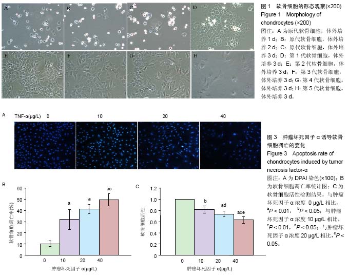
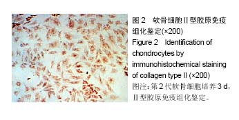
摘要
背景:
肿瘤坏死因子α是诱导骨关节炎软骨细胞凋亡的主要细胞因子,对骨关节炎的病理进程具有重要调节作用。
目的:比较不同质量浓度肿瘤坏死因子α诱导大鼠软骨细胞凋亡模型,为进一步探讨药物对软骨细胞凋亡的调控作用提供理论支持。
方法:清洁级4周龄SD大鼠,采用机械-Ⅱ型胶原酶消化法获取膝关节软骨细胞,并用不同质量浓度的肿瘤坏死因子α(0,10,20,30,40 μg/L)诱导建立软骨细胞的凋亡模型,倒置相差显微镜观察软骨细胞的形态结构,Ⅱ型胶原免疫组化鉴定软骨细胞,MTT检测软骨细胞的活性,DAPI检测软骨细胞的凋亡情况。
结果与结论:①体外培养软骨细胞,Ⅱ型胶原免疫组化染色可见胞浆呈棕黄色,为阳性细胞;②软骨细胞凋亡率,干预48 h肿瘤坏死因子α 10,20,30 μg/L明显高于0 μg/L(P < 0.01),肿瘤坏死因子α 40 μg/L明显高于10 μg/L(P < 0.01);③软骨细胞活性,干预48 h肿瘤坏死因子α 10,20,40 μg/L明显低于 0 μg/L(P < 0.01),肿瘤坏死因子α 20 μg/L明显低于10 μg/L(P < 0.05),肿瘤坏死因子α 40 μg/L明显低于10 μg/L(P < 0.01)、20 μg/L(P < 0.05)。④结果说明,20 μg/L肿瘤坏死因子α能成功建立炎症介导软骨细胞凋亡模型。

中国组织工程研究杂志出版内容重点:组织构建;骨细胞;软骨细胞;细胞培养;成纤维细胞;血管内皮细胞;骨质疏松组织工程
ORCID: 0000-0002-9723-0712(陈后煌)

关键词: 组织构建, 软骨细胞, 肿瘤坏死因子α, MTT, DAPI, Ⅱ型胶原鉴定, 凋亡模型, 炎症, 国家自然科学

Abstract:

Abstract
BACKGROUND:
Tumor necrosis factor-α (TNF-α), a main cytokine inducing chondrocyte apoptosis of osteoarthritis, plays a regulatory role in the process of osteoarthritis.
OBJECTIVE: To compare the rat models of chondrocyte apoptosis induced by different mass concentrations of TNF-α, thus providing theoretical basis for further study on the regulation of drugs on chondrocyte apoptosis.
METHODS: Chondrocytes were isolated from the knee cartilage of 4-week-old Sprague-Dawley rats of clean grade by mechanical ll collagenase digestion and were then incubated with different mass concentrations of TNF-α to induce apoptosis. The morphology of chondrocytes was observed under inverted phase contrast microscope, cells were identified using immunohistochemical staining of type II collagen, as well as the cell viability and apoptosis were detected by MTT and DAPI, respectively.
RESULTS AND CONCLUSION: (1) In vitro, the cytoplasm of chondrocytes was stained brown-yellow by using immunohistochemical staining of type II collagen. (2) At 48 hours, the apoptosis rate of chondrocytes induced by 10, 20 and 30 μg/L TNF-α was significantly higher than that of the 0 μg/L TNF-α (P < 0.01), and the apoptosis rate of chondrocytes induced by 40 μg/L TNF-α was significantly higher than that of the 10 μg/L TNF-α (P < 0.01). (3) The viability of chondrocytes induced by 10, 20 and 40 μg/L TNF-α was significantly lower than that of the 0 μg/L TNF-α (P < 0.01). In detail, the viability of chondrocytes induced by 20 μg/L TNF-α was lower than that of the 10 μg/L TNF-α (P < 0.05); the viability of chondrocytes induced by 40 μg/L TNF-α was significantly lower than that of the 10 and 20 μg/L TNF-α (P < 0.01, P < 0.05). (4) These results suggest that 20 μg/L TNF-α can successfully induce the chondrocyte apoptosis model.

中国组织工程研究杂志出版内容重点:组织构建;骨细胞;软骨细胞;细胞培养;成纤维细胞;血管内皮细胞;骨质疏松组织工程

Key words: Chondrocytes, Tumor Necrosis Factor-alpha, Apoptosis, Inflammation, Tissue Engineering

中图分类号: