中国组织工程研究 ›› 2017, Vol. 21 ›› Issue (2): 182-186.doi: 10.3969/j.issn.2095-4344.2017.02.004

• 组织工程骨及软骨材料 tissue-engineered bone and cartilage materials • 上一篇    下一篇

骨生物材料复合骨髓间充质干细胞异位成骨修复肋骨大段缺损

王俊钢1,李聪聪2,毛广显3,张  洁1,杨  翠1
  

  1. 郑州大学附属医院/南阳市中心医院,1胸外科,2特需病房一病区,河南省南阳市  473000;3北京大学附属深圳医院胸外科,广东省深圳市  518036
  • 收稿日期:2016-10-26 出版日期:2017-01-18 发布日期:2017-02-27
  • 作者简介:王俊钢,男,1978年生,河南省南阳市人,主治医师,主要从事胸外科方面疾病的研究与治疗研究。
  • 基金资助:

    深圳市科技计划项目(JCYJ20140415162338820);深圳市医疗卫生科研项目(201302068)

Bone biomaterial composited with human bone marrow mesenchymal stem cells for large costal defects

Wang Jun-gang1, Li Cong-cong2, Mao Guang-xian3, Zhang Jie1, Yang Cui1
  

  1. 1Department of Thoracic Surgery, 2First Special Ward, Affiliated Hospital of Zhengzhou University & Nanyang City Central Hospital, Nanyang 473000, Henan Province, China; 3Department of Thoracic Surgery, Peking University Shenzhen Hospital, Shenzhen 518036, Guangdong Province, China
  • Received:2016-10-26 Online:2017-01-18 Published:2017-02-27
  • About author:Wang Jun-gang, Attending physician, Department of Thoracic Surgery, Affiliated Hospital of Zhengzhou University & Nanyang City Central Hospital, Nanyang 473000, Henan Province, China
  • Supported by:

    the Science and Technology Program of Shenzhen, No. JCYJ20140415162338820; the Project of Medical and Health Research of Shenzhen, No. 201302068

摘要:

文章快速阅读:


文题释义:
海藻酸盐
:是目前生物材料中一类研究较为热点的无机物,具有可塑性强、可降解、组织低排异度及X射线透视不显影的优势,因此选取海藻酸盐作为此次实验的主要生物无活性组织。为探究改善目前治疗大段肋骨缺损的手段,实验拟采取大鼠骨髓间充质干细胞为种子细胞,在复合海藻酸盐的支撑下,结合体内及体外生物学实验,初步探究骨生物材料复合大鼠骨髓间充质干细胞异位成骨治疗肋骨大段缺损的效果。
间充质干细胞:是机体内与成骨相关的关键细胞,起源于胚胎中胚层,随着胚胎的逐渐发育,间充质干细胞可分布于机体各大间质组织中,其中以骨髓、脐血最为丰富。在机体发育过程中,间充质干细胞接受体内信号刺激后,动员开始分化并成骨,同样此类行为在创伤中更为显著。

背景:组织工程生物材料具有与自体组织相似的结构与功能。
目的:观察骨生物材料复合大鼠骨髓间充质干细胞异位成骨治疗肋骨大段缺损的效果。
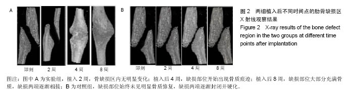
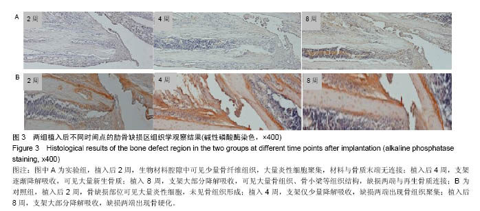
方法:取Wistar大鼠50只,制备右侧大段肋骨缺损模型,随机分为2组,对照组于骨缺损处置入氯化钙-藻酸钠凝胶,实验组于骨缺损处植入氯化钙-藻酸钠-骨髓间充质干细胞复合物。植入后2,4,8周,进行胸部X射线及骨缺损处组织形态学观察。
结果与结论:①X射线观察结果:植入后2周,实验组骨缺损区无明显变化;植入后4周,缺损部位开始出现骨质痕迹;植入后8周,缺损部位大部分充满骨质,缺损两端逐渐相接。对照组缺损部位始终未见明显骨质修复,缺损两端逐渐封闭并硬化;②骨缺损处组织形态学观察结果:植入后2周,实验组材料腔隙中可见少量骨纤维组织,大量炎性细胞聚集,材料与骨质末端无连接;对照组骨缺部位可见大量炎性细胞,未见骨组织形成。植入后4周,实验组支架逐渐降解,可见大量新生骨质;对照组支架仅少量降解,缺损两端出现骨组织聚集。植入后8周,实验组支架大部分降解,可见大量骨组织、骨小梁等组织结构,缺损两端与再生骨质连接;对照组支架大部分降解,缺损两端出现骨硬化;③结果表明:骨生物材料复合大鼠骨髓间充质干细胞可促进肋骨大段缺损的修复。

关键词: 生物材料, 骨生物材料, 海藻酸钠, 间充质干细胞, 组织工程骨, 肋骨大段缺损

Abstract:

BACKGROUND: Tissue-engineered biomaterials have the similar structure and function with autologous tissues.
OBJECTIVE: To explore the osteoinduction of the bone biomaterial composited with rat bone marrow mesenchymal stem cells in the treatment of large costal defects.
METHODS: Forty Wistar rats were enrolled used for the preparation of right large costal defect models, and then randomized into two groups, followed by the implantation of calcium chloride-sodium alginate gel (control group) or chloride-sodium alginate-bone marrow mesenchymal stem cells (experimental group). At 2, 4 and 8 weeks after implantation, chest X-ray radiograph and histological examination of the defect region were conducted.
RESULTS AND CONCLUSION: X-ray showed that in the experimental group, the defect area had no significant changes at the 2nd week after implantation until the formation of few bones at the 4th week; and at the 8th week, both ends of the defect region gradually connected, and newly formed bones were full of the defect. In contrast, the defect region in the control group showed no obvious bone healing, and both ends of the defect closed and osteosclerosis occurred. In the experimental group, there were a small amount of fibrous tissues and numerous inflammatory cells infiltratied in the material compartment, and no connection occured between the material and broken ends; there were numerous inflammatory cells but no bone tissues in the control group at the 2nd week. At the 4th week, the scaffold degraded gradually and abundant bone tissues were seen in the experimental group; the scaffold degraded little, and bone tissues aggregatied at the both defect ends in the control group. Up to the 8th week, the two kinds of scaffolds degraded mostly. A large number of bone tissues and trabeculae formed and the both defect ends were connected with the newly formed bones in the experimental groups, while in the control group, osteosclerosis appeared at both ends of the defect. To conclude, the bone biomaterial composited with rat bone marrow mesenchymal stem cells promotes the repair of large costal defects.

Key words: Alginates, Mesenchymal Stem Cells, Tissue Engineering

中图分类号: