中国组织工程研究 ›› 2016, Vol. 20 ›› Issue (24): 3605-3608.doi: 10.3969/j.issn.2095-4344.2016.24.016
• 肌肉肌腱韧带组织构建 tissue construction of the muscle, tendon and ligament • 上一篇 下一篇
前交叉韧带断裂和重建的临床流行病学分析
陈连旭,付立功
- 清华大学医学中心北京清华长庚医院骨科,北京市 102218
Clinical epidemiological study on anterior cruciate ligament rupture and reconstruction
Chen Lian-xu, Fu Li-gong
- Department of Orthopedics, Beijing Tsinghua Chang Gung Hospital, Medial Center, Tsinghua University, Beijing 102218, China
摘要:
文章快速阅读:
.jpg) 文题释义:
前交叉韧带:膝关节内重要的韧带之一,起自股骨外侧髁内侧面,止于胫骨髁间嵴前部,主要维护膝关节的前向稳定性和旋转稳定性。前交叉韧带在解剖和功能上分为前内束和后外束,前内束主要负责前向稳定性,后外束主要负责旋转稳定性。前内束和后外束在屈膝时呈交叉状态,伸直时呈平行状态。
前交叉韧带解剖重建:是前交叉韧带重建最新的重建方法,通过前交叉韧带的原止点重建,恢复原有前交叉韧带的止点位置、走形方向和韧带大小,完全恢复前交叉韧带的功能。解剖重建的关键是骨道定位,精髓是交叉韧带止点残端中央定位。解剖重建包括解剖单束重建、解剖双束重建、前内束和后外束解剖部分重建。
文题释义:
前交叉韧带:膝关节内重要的韧带之一,起自股骨外侧髁内侧面,止于胫骨髁间嵴前部,主要维护膝关节的前向稳定性和旋转稳定性。前交叉韧带在解剖和功能上分为前内束和后外束,前内束主要负责前向稳定性,后外束主要负责旋转稳定性。前内束和后外束在屈膝时呈交叉状态,伸直时呈平行状态。
前交叉韧带解剖重建:是前交叉韧带重建最新的重建方法,通过前交叉韧带的原止点重建,恢复原有前交叉韧带的止点位置、走形方向和韧带大小,完全恢复前交叉韧带的功能。解剖重建的关键是骨道定位,精髓是交叉韧带止点残端中央定位。解剖重建包括解剖单束重建、解剖双束重建、前内束和后外束解剖部分重建。
文题释义:
前交叉韧带:膝关节内重要的韧带之一,起自股骨外侧髁内侧面,止于胫骨髁间嵴前部,主要维护膝关节的前向稳定性和旋转稳定性。前交叉韧带在解剖和功能上分为前内束和后外束,前内束主要负责前向稳定性,后外束主要负责旋转稳定性。前内束和后外束在屈膝时呈交叉状态,伸直时呈平行状态。
前交叉韧带解剖重建:是前交叉韧带重建最新的重建方法,通过前交叉韧带的原止点重建,恢复原有前交叉韧带的止点位置、走形方向和韧带大小,完全恢复前交叉韧带的功能。解剖重建的关键是骨道定位,精髓是交叉韧带止点残端中央定位。解剖重建包括解剖单束重建、解剖双束重建、前内束和后外束解剖部分重建。摘要
背景:前交叉韧带解剖重建是治疗前交叉韧带断裂的主要方法。目前针对前交叉韧带重建,从组织胚胎、解剖结构和生物力学,到重建材料、操作技术和重建后康复都进行了全面细致的研究,但缺乏前交叉韧带断裂和重建的临床流行病学资料。
目的:分析前交叉韧带断裂和重建的临床流行病学特点,为预防和治疗前交叉韧带断裂提供指导。
方法:统计了前交叉韧带断裂重建患者352例,对患者的年龄、性别、受伤原因、受伤机制、就诊时间和前交叉韧带断裂后对半月板和关节软骨的影响,以及手术过程中的发现情况、手术方式和重建材料等进行分析。
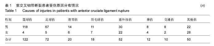
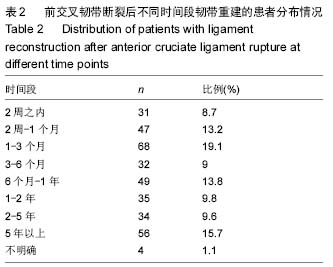
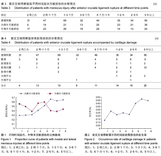
结果与结论:①前交叉韧带断裂多发生于男性年轻人,左膝多于右膝;②男性患者多发生于篮球、足球和意外伤,女性患者多发生于意外伤、羽毛球和滑雪伤,损伤机制以膝关节内旋外翻伤多见;③手术时间以前交叉韧带断裂伤1-3个月多见,常伴有半月板和关节软骨损伤。外侧半月板损伤发生率相对稳定,内侧半月板损伤在前交叉韧带断裂超过半年后明显增多。关节软骨损伤以髌骨软骨损伤为主,超过1年,内侧髁软骨损伤显著增加;④韧带重建方式以解剖单束重建为主,骨道定位可参考住院医师嵴和束间嵴,重建材料多为自体半腱股薄肌肌腱;⑤结果提示,前交叉韧带断裂应早期进行前交叉韧带的解剖重建,恢复膝关节的稳定性,防止内侧半月板和股骨内侧髁软骨的继发损伤。
中国组织工程研究杂志出版内容重点:组织构建;骨细胞;软骨细胞;细胞培养;成纤维细胞;血管内皮细胞;骨质疏松;组织工程
ORCID: 0000-0003-3917-991X(陈连旭)
中图分类号:
.jpg) 文题释义:
前交叉韧带:膝关节内重要的韧带之一,起自股骨外侧髁内侧面,止于胫骨髁间嵴前部,主要维护膝关节的前向稳定性和旋转稳定性。前交叉韧带在解剖和功能上分为前内束和后外束,前内束主要负责前向稳定性,后外束主要负责旋转稳定性。前内束和后外束在屈膝时呈交叉状态,伸直时呈平行状态。
前交叉韧带解剖重建:是前交叉韧带重建最新的重建方法,通过前交叉韧带的原止点重建,恢复原有前交叉韧带的止点位置、走形方向和韧带大小,完全恢复前交叉韧带的功能。解剖重建的关键是骨道定位,精髓是交叉韧带止点残端中央定位。解剖重建包括解剖单束重建、解剖双束重建、前内束和后外束解剖部分重建。
文题释义:
前交叉韧带:膝关节内重要的韧带之一,起自股骨外侧髁内侧面,止于胫骨髁间嵴前部,主要维护膝关节的前向稳定性和旋转稳定性。前交叉韧带在解剖和功能上分为前内束和后外束,前内束主要负责前向稳定性,后外束主要负责旋转稳定性。前内束和后外束在屈膝时呈交叉状态,伸直时呈平行状态。
前交叉韧带解剖重建:是前交叉韧带重建最新的重建方法,通过前交叉韧带的原止点重建,恢复原有前交叉韧带的止点位置、走形方向和韧带大小,完全恢复前交叉韧带的功能。解剖重建的关键是骨道定位,精髓是交叉韧带止点残端中央定位。解剖重建包括解剖单束重建、解剖双束重建、前内束和后外束解剖部分重建。