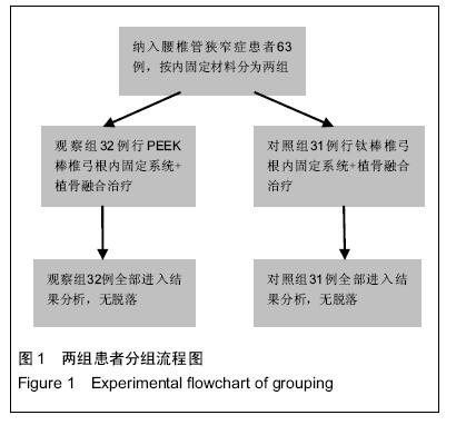
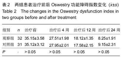
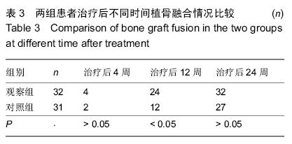
| [1] 王金辉.2种TLIF手术方法治疗腰椎滑脱合并椎管狭窄症的疗效比较[J].中国伤残医学,2015,23(9):44-45.[2] 田地,赵伟.植骨融合术在治疗退变性腰椎管狭窄症中的应用进展[J].医学综述,2014,20(19):3549-3551.[3] 刘培盛,刘小臻,乔雪静.椎间融合器植入与单纯自体颗粒骨植骨治疗退变性腰椎管狭窄症的比较[J].中华医学杂志,2014,(35):2731-2735.[4] 傅智轶,史建刚,贾连顺.有限椎板切除减压与全椎板切除减压内固定术治疗腰椎管狭窄症的疗效对比[J].中国矫形外科杂志,2014,22(15):1347-1352.[5] 陈铭,范海泉,江洋,等.退变性腰椎侧凸合并腰椎管狭窄症患者的个体化手术治疗[J].中国骨与关节杂志,2014, 13(12):931-934.[6] 李德平,毛波,潘伟明,等.半椎板减压联合椎间植骨融合和单侧椎弓根内固定治疗腰椎管狭窄症伴退变性Ⅰ度滑脱34例疗效观察[J].海南医学,2014,25(18):2747-2749.[7] 黄东永,黄远源,陈敏,等.腰椎后路减压内固定融合术(PLIF)治疗退行性腰椎管狭窄症(DLSS)的临床效果[J].齐齐哈尔医学院学报,2014,31(15):2252-2253.[8] 张亚峰,杨惠林,唐天驷,等.后路椎体间融合术后融合器脱出的原因及其翻修术[J].中国脊柱脊髓杂志,2006,16(12): 909-912.[9] 江伟,叶蜀新.PEEK Cage PTLIF治疗退变性腰椎不稳及腰椎管狭窄症[C].//四川省医学会第十四次骨科学术会议论文集,2010:75.[10] 钱邦平,唐天驷,杨同其,等.后路椎体间融合治疗严重腰椎管狭窄症初步报告[J].中国脊柱脊髓杂志,2002,12(5): 371-373.[11] 王玉松,赵筑川,罗春山,等.腰椎后路固定联合PEEK椎间融合治疗退变性腰椎管狭窄症的疗效分析[C].//第25届全国脊柱脊髓学术会议暨2013年贵州省骨科年会论文集,2013:158.[12] 朱建炜,刘璠,张烽,等.腰椎间孔入路腰椎体间植骨融合结合椎弓根螺钉置入内固定:能提高退行性病变腰椎的稳定性与植骨融合率吗[J].中国组织工程研究与临床康复, 2010,14(13):2425-2428.[13] 史瑞明,李国胜,张义峰,等.钉棒置入与椎间融合修复极外侧型腰椎间盘突出症:远期腰椎稳定性随访[J].中国组织工程研究,2014,18(40):6464-6470.[14] 樊友亮,吴一雄,胡辉东,等.经椎弓根动态稳定系统(Dynesys)与后路椎间融合治疗腰椎管狭窄症的临床比较研究[J].中国骨与关节损伤杂志,2013,28(11): 1016-1019.[15] 杨红伟,陈永飞.椎间减压、椎弓根半裸置钉内固定加植骨术治疗腰椎滑脱症合并椎管狭窄34例[J].广西医科大学学报,2011,28(1):115-117.[16] 樊友亮.经椎弓根动态稳定系统(Dynesys)与后路椎间融合治疗腰椎管狭窄症的临床比较研究[D].苏州大学, 2012.[17] 曹俊明,张迪,申勇,等.椎间融合器植入并双侧下关节突切除治疗退变性腰椎管狭窄症[J].中国组织工程研究与临床康复,2010,14(17):3226-3230.[18] 吴大鹏,刘晓谭,徐海斌.X-Tube辅助下微创经椎间孔腰椎间融合术治疗退变性腰椎管狭窄症[J].中国现代医学杂志, 2014,24(21):54-59.[19] 崔后春,王汝渔,荆鑫.个体化椎板减压结合椎间融合或后外侧融合内固定治疗退变性腰椎管狭窄症[J].中国骨与关节损伤杂志,2014,29(9):932-933.[20] 文天林,刘秀梅,杜培.Quadrant微创通道下单侧开窗减压与开放减压内固定术治疗退变性腰椎管狭窄症病例对照研究[J].中国骨伤,2014,28(8):658-662.[21] 丁辉耀,李绍波,朱斌,等.短节段内固定联合单枚椎间融合器治疗腰椎管狭窄症患者的疗效观察[J].中国民康医学, 2015,27(6):1-4.[22] 贾希瑞,刘洋,赵立新.电针配合腰背肌锻炼治疗黄韧带肥厚型腰椎管狭窄症临床观察及治疗体会[J].光明中医, 2015,30(2):317-318.[23] 巴根,贾长青.老年退变性腰椎管狭窄症的个体化手术治疗及临床疗效观察[J].东南大学学报:医学版,2015,56(1): 22-26.[24] 勾旭升,陈立民,张捍军,等.腰椎管狭窄症脊椎融合术适应症研究进展[J].现代生物医学进展,2015,15(3):581-583.[25] 江铭,朱文雄,吴海谊.PLIF手术治疗退行性腰椎管狭窄症伴腰椎失稳的疗效观察[J].右江民族医学院学报,2015, 37(1):68-69.[26] 韩利伟.后路椎间盘镜手术治疗老年腰椎管狭窄症的临床效果分析[J].中国现代药物应用,2015,9(4):30-31.[27] 乐锦波,杜远立.半椎板切除减压术与全椎板切除椎间融合内固定术治疗腰椎管狭窄症的疗效比较[J].实用医院临床杂志,2014,11(5):199-200.[28] 梁昌详,昌耘冰,沈梓维,等.椎管减压棘突间Coflex置入术治疗L4/5退变性腰椎管狭窄症的5年随访结果[J].中国脊柱脊髓杂志,2014,24(12):1072-1078.[29] 代晓平.椎弓根内固定与椎间融合器植骨融合治疗腰椎管狭窄症疗效观察[J].现代中西医结合杂志,2014, 23(31): 3488-3490.[30] 陈新宇.有限减压、固定、融合术在退行性腰椎管狭窄症治疗中的应用[J].中国基层医药, 2014,21(16):2449- 2451.[31] Barz T, Melloh M, Staub LP, et al. Increased intraoperative epidural pressure in lumbar spinal stenosis patients with a positive nerve root sedimentation sign. Eur Spine J. 2014;23(5):985-990.[32] Arai Y, Hirai T, Yoshii T, et al. A prospective comparative study of 2 minimally invasive decompression procedures for lumbar spinal canal stenosis: Unilateral laminotomy for bilateral decompression (ULBD) versus muscle-preserving interlaminar decompression (MILD).Spine. 2014;39(4): 332-340.[33] Tsutsui S, Kagotani R, Yamada H, et al. Can decompression surgery relieve low back pain in patients with lumbar spinal stenosis combined with degenerative lumbar scoliosis? Eur Spine J. 2013; 22(9):2010-2014.[34] 徐浩.新型单枚斜向植入腰椎椎间融合器的生物力学及临床研究[D].第二军医大学,2009.[35] 葛钟牛.PEEK与钛金属椎弓根内固定系统联合椎间植骨融合治疗腰椎管狭窄症的疗效对比[D].山东中医药大学, 2012. |
.jpg)

.jpg)
.jpg)