中国组织工程研究 ›› 2016, Vol. 20 ›› Issue (9): 1221-1226.doi: 10.3969/j.issn.2095-4344.2016.09.001

• 人工假体 artificial prosthesis •    下一篇

股骨内髁参数对单髁置换中截骨及假体设计的参考意义

祁家龙1,尹宗生1,马广文2   

  1. 1安徽医科大学第一附属医院关节外科中心,安徽省合肥市 230000;2安徽医科大学第四附属医院骨科,安徽省合肥市 230000
  • 收稿日期:2016-01-23 出版日期:2016-02-19 发布日期:2016-02-19
  • 作者简介:祁家龙,男,1989年生,汉族,安徽医科大学在读硕士。

Significance of femoral condyle parameters in osteotomy in unicompartmental knee arthroplasty and prosthesis design

Qi Jia-long1, Yin Zong-sheng1, Ma Guang-wen2   

  1. 1Joint Surgery Center, First Affiliated Hospital, Anhui Medical University, Hefei 230000, Anhui Province, China; 2Department of Orthopedics, Fourth Affiliated Hospital, Anhui Medical University, Hefei 230000, Anhui Province, China
  • Received:2016-01-23 Online:2016-02-19 Published:2016-02-19
  • About author:Qi Jia-long, Studying for master’s degree, Joint Surgery Center, First Affiliated Hospital, Anhui Medical University, Hefei 230000, Anhui Province, China

摘要:

文章快速阅读:

文题释义:

股骨内髁参数:文中指股骨内髁在三维空间里的大小,股骨内髁在某些层面上近似圆形(相关文献报道里已形成某种共识),具体通过对内髁上特征性的俩点所确定的圆弧进行测量获得直径大小,文中所测3组数据及其相关性直接决定了单髁置换假体的尺寸。

个性化截骨:为针对个人差异采用的特殊截骨方式,膝关节大小存在个体差异性,但是假体尺寸相对固定,选择可能性小,会存在一定程度的不完全匹配,个性化截骨能够获得更好的假体匹配。


背景:膝关节参数测量对于研究和设计适合国人的假体有着重要的意义,国内目前对全膝关节假体研究较多,单髁假体参数化测量较少。
目的:获得国人正常股骨内髁参数,探讨其对单髁置换过程中截骨及假体设计的意义。
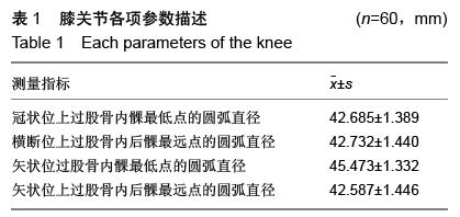
方法:选取60例60膝国人正常膝关节,利用薄层CT平扫及后处理技术测量冠状位上过股骨内髁最低点的圆弧直径、横断位上过股骨内后髁最远点的圆弧直径、矢状位上过股骨内后髁最远点的圆弧直径及矢状位上过股骨内髁最低点的圆弧直径,并分析其与性别、身高的关系。
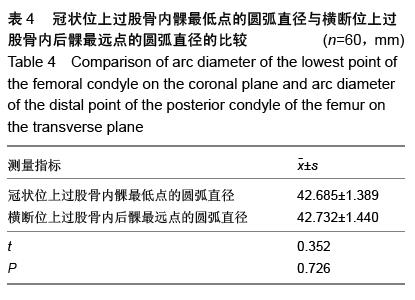
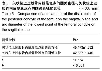
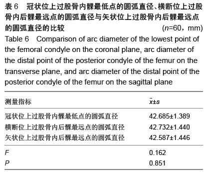
结果与结论:测得所有志愿者的冠状位上过股骨内髁最低点的圆弧直径的平均值为(42.685±1.389) mm、横断位上过股骨内后髁最远点的圆弧直径的平均值为(42.732±1.440) mm、矢状位上过股骨内髁最低点圆弧直径的平均值为(45.473±1.332) mm、矢状位上过股骨内后髁最远点的圆弧直径的平均值为(42.587±1.446) mm。结果显示,膝关节内髁相关参数与身高呈正相关,男性明显大于女性。矢状位上过股骨内髁最低点的圆弧直径显著大于过股骨内后髁最远点的圆弧直径(P < 0.001);冠状位上过股骨内髁最低点的圆弧直径、横断位上过股骨内后髁最远点的圆弧直径、矢状位上过股骨内后髁最远点的圆弧直径三者无明显差异。 

ORCID: 0000-0002-6042-613X(祁家龙)

关键词: 骨科植入物, 人工假体, 单髁置换, 股骨内髁参数, 股骨内髁最低端, 股骨内后髁最远点, 内髁截骨

Abstract:

BACKGROUND: Knee-parameter measurements play an important role in the designing of the knee prosthesis. Currently, we have more and more research of the total knee replacement, while uni-condylar knee replacement study is few. 
OBJECTIVE: To obtain the parameters of the normal femoral condyles and explore its correlation with osteotomy and prosthesis design of the knee joint during uni-condylar knee replacement.
METHODS: Normal knee joints of 60 cases (60 knees) were selected. We measured the parameters by using thin-section CT scan and post-processing techniques, including arc diameter of the lowest point of the femoral condyle on the coronal plane, arc diameter of the distal point of the posterior condyle of the femur on the transverse plane, arc diameter of the distal point of the posterior condyle of the femur on the sagittal plane, and arc diameter of the lowest point of the femoral condyle on the sagittal plane, and analyzed the correlation with sex and height. 
RESULTS AND CONCLUSION: The diameter of the arc that passes through the lowest point of femoral medial condyle in the coronal plane was (42.685±1.389) mm. The diameter of the arc that passes through the farthest point of posterior of femoral medial condyle in the cross-section was (42.732±1.440) mm. The diameter of the arc that passes through the lowest point of femoral medial condyle on the sagittal plane was (45.473±1.332) mm. The diameter of the arc that passes through the farthest point of posterior of femoral medial condyle on the sagittal plane was (42.587±1.446) mm. The results illustrate that knee condyle related parameters were positively correlated with height. Parameters in males were significantly greater than in females. The diameter of the arc that passes through the lowest point of femoral medial condyle on the sagittal plane was significantly larger than that of the farthest point of femoral posterior medial condyle on the sagittal plane (P < 0.001). There was no significant difference among the diameter of the arc that passes through the lowest point of femoral medial condyle on the coronal plane, the diameter of the arc that passes through the farthest point of posterior of femoral medial condyle on the cross-section and that of the farthest point of femoral posterior medial condyle on the sagittal plane.