| [1] 王继荣,刘成环,孙晓芹.强直性脊柱炎骶髂关节的CT诊断价值[J].青海医药杂志,2013,43(2):73-75.
[2] 俞永梅,徐亮,张锡龙,等,X线,CT和MRI在强直性脊柱炎骶髂关节病变中的诊断价值[J].皖南医学院学报,2013,5(1):404-407.
[3] 朱利君,王利伟,钱少圭,等,磁共振诊断强直性脊柱炎骶髂关节病变的价值[J].中国CT和MRI杂志,2012,10(5):86-88.
[4] Mager AK, Althoff CE, Sieper J, et al. Role of whole body magnetic resonance imaging in diagnosing early spondyloarthritis.Eur J Radio.2009; 71:182-188.
[5] Wittram C,Whitchouse GH. Nomal variation in the magnetic resonance imaging appearances of the sacroiliac joints pitfalls in the diagnosis of sacroliitis.Clin Radiol.1995; 50(6):371-376.
[6] Modl JM, Sether LA, Haughton VM, et al.Articular cartilage: correlation of histologic zones with signal intensity at MR imaging, Radiology.1991; 181:853-855.
[7] loeuille D,Olivier P,Watrin A,et al.The biochemical content of articular cartilage:an original MRI approach.Biorheology. 2002; 39(1-2):269-276.
[8] 黄振国,张雪哲,洪闻,等.早期强直性脊柱炎骶髂关节病变的X线、CT和MRI对比研究[J].中华放射学杂志,2011,45(11):1040-1044.
[9] Ganz R,Leunig M,Leunig-Ganz K,et al.Theetiology of osteoar th r itis of the hip:integ rated mechanical concept. Clinorthop. 2008; 466(2):264-272.
[10] 刘丙木,王淑梅,崔建岭,等,10-20岁健康志愿者骶髂关节软骨MRI序列对照研究[J].临床放射学杂志,2008,2(12):1786-1789.
[11] Marzo-Ortega H, McGonagle D, Bennett AN. Magnetic resonance imaging in spondyloarthritis.Curr Opin Rheumatol. 2010;22(4):381-387.
[12] Malaviya AP, Ostor AJ.Early diagnosis crucial in ankylosing spondylitis. Practitioner.2011;255(1746):21-24.
[13] Vander Cruyssen B, Munoz-Gomariz E, Foni P, et al .Hip involve menin ankylosing spondylifs:epide miology and risk factors associated with hip replace ment surgery. Rheumatology(Oxtord).2010;49(1):73-81.
[14] Ai F,Ai T,Li X,et al. value of diffusion-weighted magnetic resonance imaging in early diagnosis of ankylosing spondylitis. Rheumatol Int.2012;32(12):4005-4013.
[15] Sanal HT, Yilmaz S, Kalyoncu U, et al. alvalue of DWI in visual assessment of activity of sacroiliitis in longstanding ankylosing spondylitis patients.Skeletal Radiol.2013;42(2):289-293.
[16] 艾飞,田丹,张炜,等.磁共振弥散加权成像诊断早期强直性脊柱炎的价值[J].中华医学杂志,2013,93(11):811-815.
[17] 俎金燕,王晨光,贾宁阳,等.腰椎间盘退行性变的磁共振弥散加权成像研究[J].实用放射学杂志,2013,29(4):611-614.
[18] Pan C,Hu DY,Zhang W,et al. Role of diffusion-weighted imaging in early ankylosing spondylitis.Chin Med J(Engl). 2013;126(4):668-673.
[19] Dallaudiere B,Dautry R,Preux PM,et al.comparison of apparent diffusion coefficient in spondylarthritis axial active inflammatory Lesions and type 1 Modic changes.Eur J Radiol. 2014;83(2):366-370.
[20] 丁庆国,贾传海,陆永明,等.振对强直性MRI联合DWI在强直性脊柱炎诊断中的价值,[J].临床放射学杂志,2012,31(5):693-696.
[21] 冯勋树,张俊湖,杨君.急性期脑梗死患者弥散加权成像皮质层状坏死表现的临床意义[J].中华行为医学与脑科学杂志, 2012, 21(3):255-257.
[22] 王健,姚秀忠,饶圣祥,等.3.0T磁共振弥散加权成像在胰腺癌中的应用价值[J].医学影像学杂志,2012,22(1):91-93.
[23] Baraliakos X, Braun J.Hip involve ment in ankylosing spondylitis:what is the verdict Rheumatology(Oxtord). 2010; 49(1):3-4. |
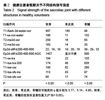
.jpg)
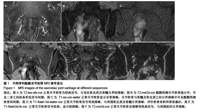
.jpg)