In early period, Wilkinson[17] pointed out that human acetabulum shares similarity with rabbit. Originally, people would drag lower extremity with excessive physical exertion to get DDH animal models while now it has been evolved into using plastic cannula, intramedullary needle and percutaneous fixation to straighten and fix the knee joint[8]. In this experiment, these young rabbits were fixed with plaster fixation and with extended knee joint, so their hips were under mechanical stress of muscle skeleton under a long time and finally got the DDH models successfully, this has been provided a theoretical foundation on the importance of the acquired environment effect on this disease as well as proved that DDH is a “developmental” disease to some extent.
DDH refers to children of different ages present dysplasia of hip joint in various forms, which is the most common disease of pediatric orthopedics. Mild DDH causes high morbidity of osteoarthritis for grown up. It is reported that 76% hip joint arthritis is caused by DDH, most of them have to take total hip replacement before the age of 50[18]. Normal chondrocytes secrete proteoglycan and show prunosus after staining with toluidine blue. After compressed, the invisible layer of articular cartilage surface having immunologic barrier to type II collagen antibody will be destroyed, then collagen fibers expose in synovial and cause chondrocytes damages as well as dysfunction and secret abnormal proteoglycans, therefore toluidine blue fails staining or with unevenly staining. In this experiment, it is observed that more visible fibrosis in many chondrocytes of surface layer, chaotic arrangement of cells in the middle layer, cells in columnar cell layer reduction as well as disordered, uneven toluidine blue staining and failed staining on matrix, which indicates that chondrocytes became degenerated due to compression in early DDH and then caused ECM elements changes in quantity and quality such as proteoglycans. This may be the pathological basis of osteoarthritis in prophase.
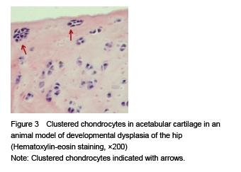
A lot of clustered chondrocytes in articular cartilage are the pathological feature for osteoarthritis with cartilaginous degeneration; as for mild osteoarthritis, metabolism of chondrocyte is active thus cells will be clustered. Proliferation of chondrocyte induces chondrocyte close to surface layer to cluster in early stage of osteoarthritis[19]. Local clustered chondrocytes in this experiment verifies that uneven stress of cartilage indifferent areas in early stage of DDH will affect the mechanical biological environment of chondrocytes.
Aigner et al [15] found that there was an obvious proliferation of chondrocytes in osteoarthritis; and presumed that cartilage degeneration in osteoarthritis started from macromolecule protein synthesis changes, which reflected as synthesis changes of type II collagen protein and proteolytic enzyme. In 1990, the first genetic mutation responsible for osteoarthritis was reported after researchers found a high rate of osteoarthritis with a mild chondrodysplasia in the family of a 44-year-old patient with degenerative changes of both hips. The mutation responsible for osteoarthritis in this family occurred in the type II collagen gene, COL2A1 (collagen, type II, alpha 1), which encodes a protein expressed almost exclusively in cartilage[20-21]. This mutation weakens the matrix and leads to premature degeneration of the cartilage[22].
Although a healthy adult’s articular cartilage has rich type II collagen, only little type II collagen mRNA expression in chondrocytes, which proves that anabolism of type II collagen in normal articular chondrocytes is weak. Because the expression of type II collagen mRNA in articular chondrocytes of osteoarthritis significantly increases, it indicates that injured chondrocytes may repair ECM with the extra synthesis of collagen[23]. Cellular metabolism is active in minor injured articular cartilage, gene expression of type II collagen and proteoglycans is higher than that in the control group. With the increment of gene expression, the synthesis of type II collagen increases also and meanwhile the deliquescence and denaturation of collagen increases obviously. Functions of chondrocytes will change and compound more matrix metalloproteinases to participate in deliquescence and denaturation of type II collagen, which will destroy the balance of ECM maintained by matrix components including type II collagen. Collagen total amount in cartilage during early and middle stage of osteoarthritis is up; the increment of synthesis of type II collagen in early stage shows that type II collagen is stained darker than the normal situation, especially around the chondrocyte cells and kytoplasm, the increment of collagen is larger than that of destroyed before cartilage reach the certain damage degree[24].
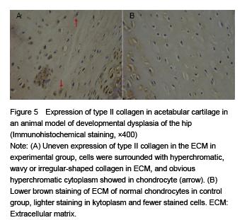
In this experiment, anachromasis of type II collagen in ECM of dysplastic acetabular cartilage suggested that more collagen in ECM; anachromasis of collagen in chondrocytes suggested that stronger synthesis of collagen. Western-blot assay detected that the expression of type II collagen protein rose significantly, further explained that when DDH acetabular cartilage under continuous abnormal stress, chondrocytes were activated and cartilaginous degeneration started then synthesized many extra cellular matrix like collagen and other macromolecule protein to adapt this kind of stress changes. Olistherozones with blooming-effect were also visible around chondrocytes in this experiment, the cause of it lay in the changes of chondrocytes function and synthesis of matrix metalloproteinases would destroy matrix components like type II collagen around cells.
Morphological change of acetabular cartilage and abnormal expression of type II collagen in early stage of DDH model which generated under mechanical stress were observed in this experiment, suggesting that these degeneration changes may be a compensatory repair reaction of chondrocytes to environmental factors (such as the mechanical changes and cytokine). Once this compensatory repair reaction is decompensation, cartilage may occur inconvertible degeneration changes and present the syndrome of osteoarthritis. Therefore, the amount and intensity of type II collagen are probably interrelated with different cartilage degeneration degrees, furthermore, as a biological parameter that reflects cartilaginous degeneration to which degree osteoarthritis will occur has not yet studied.
.jpg)
.jpg)

.jpg)
.jpg)