中国组织工程研究 ›› 2015, Vol. 19 ›› Issue (44): 7148-7155.doi: 10.3969/j.issn.2095-4344.2015.44.018

• 骨科植入物 orthopedic implant • 上一篇    下一篇

制备幼兔骺板周围骨折模型:跨骺板钢板置入内固定影响骺板的生长发育?

崔庆达1,刘 伟2,王 鑫3,马 龙4,张世谦2,潘 琦2,毕郑刚2,耿 硕2   

  1. 1青岛市胶州中心医院关节创伤科,山东省青岛市  266300;2哈尔滨医科大学附属第一医院骨科,黑龙江省哈尔滨市  150001;3武警黑龙江省总队医院,黑龙江省哈尔滨市  150076;4解放军黑龙江省军区医院,黑龙江省哈尔滨市  150047
  • 收稿日期:2015-08-19 出版日期:2015-10-22 发布日期:2015-10-22
  • 通讯作者: 毕郑刚,博士生导师,主任医师,哈尔滨医科大学附属第一医院骨科,黑龙江省哈尔滨市 150001
  • 作者简介:崔庆达,男,1982年生,山东省兰陵县人,汉族,2011年哈尔滨医科大学毕业,硕士,主治医师,主要从事关节周围骨折损伤研究。 并列第一作者:刘伟,男,1973年生,河北省秦皇岛市人,汉族,博士,副主任医师,主要从事关节周围骨折损伤研究。
  • 基金资助:

    黑龙江省教育厅资助项目(12531370)

Preparation of peri-epiphyseal fracture models in young rabbits: cross-epiphyseal plate internal fixation affects epiphyseal growth? 

Cui Qing-da1, Liu Wei2, Wang Xin3, Ma Long4,Zhang Shi-qian2, Pan Qi2, Bi Zheng-gang2, Geng Shuo2   

  1. 1Department of Joint Trauma, Jiaozhou Central Hospital of Qingdao, Qingdao 266300, Shandong Province, China; 2Department of Orthopedics, First Affiliated Hospital of Harbin Medical University, Harbin 150001, Heilongjiang Province, China; 3Harbin Armed Police Hospital, Harbin 150076, Heilongjiang Province, China; 4PLA Military Hospital of Heilongjiang Province, Harbin 150047, Heilongjiang Province, China
  • Received:2015-08-19 Online:2015-10-22 Published:2015-10-22
  • Contact: Bi Zheng-gang, Chief physician, Doctoral supervisor, Department of Orthopedics, First Affiliated Hospital of Harbin Medical University, Harbin 150001, Heilongjiang Province, China
  • About author:Cui Qing-da, Master, Attending physician, Department of Joint Trauma, Jiaozhou Central Hospital of Qingdao, Qingdao 266300, Shandong Province, China Liu Wei, M.D., Associate chief physician, Department of Orthopedics, First Affiliated Hospital of Harbin Medical University, Harbin 150001, Heilongjiang Province, China Cui Qing-da and Liu Wei contributed equally to this work
  • Supported by:

    Femoral Fractures; Internal Fixators; Models, Animal; Mast Cells; Tissue Engineering

摘要:

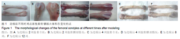
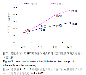
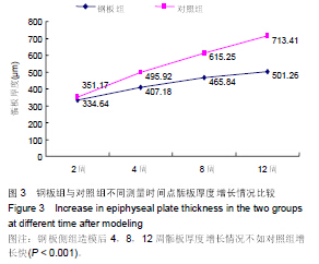
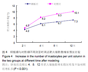
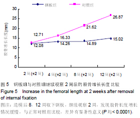
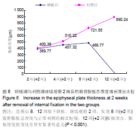
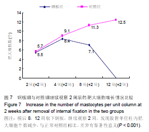
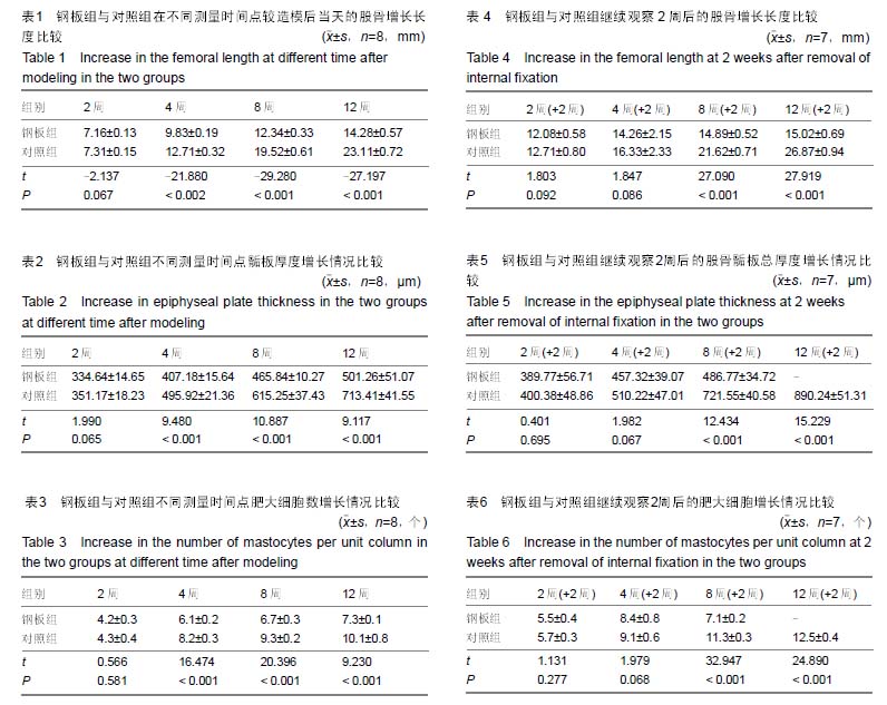
背景:当儿童四肢骨折涉及骨骺和干骺端骨折时,治疗方法多种多样。针对不同的治疗方法,对骺板生长发育影响的研究也较多。但多局限于不同直径克氏针或空心螺钉内固定对骺板发育的影响,而钢板置入跨越骺板内固定,对骺板生长发育影响及影响程度目前国内外文献报道较少。
目的:制备幼兔骺板周围骨折模型,观察跨骺板钢板置入内固定以及取出内固定物后对骺板生长发育的影响。
方法:建立幼年家兔右股骨远端骺板上方5 mm横断骨折模型60只,应用相同型号“L”型钢板和4枚螺钉跨骺板及骺板周围骨折线钢板内固定。造模后2,4,8,12周处死8只,取出股骨标本,测量股骨长度、骺板总厚度及单位柱内肥大细胞计数,观察组织形态学和特殊软骨染色中肥大细胞及骺板厚度的变化;另外7只取出内固定,继续养殖2周后处死,再次观察以上指标。左股骨远端骺板作为对照组。
结果与结论:钢板组在造模后2周上述观察指标与对照组对比,差异无显著性意义(P > 0.05)。钢板组在造模后4,8,12周上述观察指标与对照组对比,差异有显著性意义(P < 0.05或< 0.001)。造模后2,4周取下钢板,继续观察2周,发现股骨长度、骺板总厚度及单位柱内肥大细胞计数等观察指标都有不同程度恢复,与对照组比较差异无显著性意义(P > 0.05)。但造模后8,12周取下钢板,继续观察2周,发现股骨长度、骺板总厚度短缩,单位柱内肥大细胞个数明显减少,与对照组比较,差异有非常显著性意义 (P < 0.001)。提示跨骺板钢板内固定初期(2周内),在内固定物不伤及骺板的前提下,适当压应力作用对骺板生长发育未见显著影响;但持久过度钢板限制(> 4周)将导致骺板生长部分或者完全阻滞,主要引起肢体成角畸形及骺板发育阻滞。跨骺板分别固定2,4,8,12周后,取出内固定继续观察2周,发现在第2,4周两个时间点取出内固定后,股骨长度、骺板总厚度、单位柱内肥大细胞计数等观察指标都有不同程度恢复或者接近正常值。而在第8,12周两个时间点取出内固定后,继续观察2周,上述观察指标与正常值存在显著差异,说明增殖层和肥大层软骨细胞细胞核失去分化增殖能力,股骨长度和骺板厚度很难恢复到正常的程度。
中国组织工程研究杂志出版内容重点:人工关节;骨植入物;脊柱骨折;内固定;数字化骨科;组织工程

关键词: 骨科植入物, 骨植入物, 骺板周围骨折, 跨骺板, 钢板内固定, 动物实验

Abstract:

BACKGROUND: There are various therapies for children limb fractures involving the epiphysis or the metaphysis. According to the different methods, studies on the growth of the epiphyseal plate are a lot, most of which focus on 
the effects of Kirschner wires with different diameters or hollow screw internal fixation on the development of epiphyseal plate. However, there are rare studies on the influence of cross-epiphyseal plate internal fixation on the growth of epiphyseal plate as well as the influence level.
OBJECTIVE: To prepare a peri-epiphyseal fracture model in young rabbits and to observe the effects of cross-epiphyseal plate implantation and removal on the growth of epiphyseal plate.
METHODS: Traverse fracture models were made 5 mm above the right femoral distal epiphyseal plate of 60 young rabbits, and then fixed with suitable “L” steel plate and four screws across the epiphyseal plate and peri-epiphyseal fracture line. The left side served as control. Eight rabbits were killed and observed at 2, 4, 8, 12 weeks after modeling, respectively, to take out the femoral specimens for measurement of femoral length, thickness of the epiphyseal plate, and number of mastocytes per unit column. Histopathology observation was done and changes in mastocytes and thickness of the epiphyseal plate were detected. Another seven rabbits were selected to remove the metal plate, continued to feed for 2 weeks and finally executed to observe the above-mentioned indexes.
RESULTS AND CONCLUSION: (1) There were significant differences in the above indexes between the plate and control groups at 4, 8, 12 weeks after modeling (P < 0.05 or P < 0.001) but not at 2 weeks after modeling (P > 0.05). These findings indicate that within 2 weeks after cross-epiphyseal plate internal fixation, proper pressue has no remarkable influence on the growth of epiphyseal plate; but after persistent internal fixation (> 4 weeks), the growth of epiphyseal plate can be partially or completed retarded. (2) At 2 and 4 weeks after modeling, the plate was removed, and 2 weeks later, the femoral length, thickness of the epiphyseal plate and mastocyte counting per unit column were improved to different extents, and there were no differences between the plate and control group (P > 0.05). At 8 and 12 weeks after modeling, the plate was removed, and 2 weeks later, the femoral length and thickness of the epiphyseal plate were shortened, and the number of mastocytes per unit column was decreased obviously, which significantly differed from the control group (P < 0.001). These findings indicate that the chondrocytes in the proliferative and hypertrophy layers lose the differentiation and proliferation abilities, and the femoral length and epiphyseal plate thickness are difficult to recover. 
中国组织工程研究杂志出版内容重点:人工关节;骨植入物;脊柱骨折;内固定;数字化骨科;组织工程

Key words: Femoral Fractures, Internal Fixators, Models, Animal, Mast Cells, Tissue Engineering