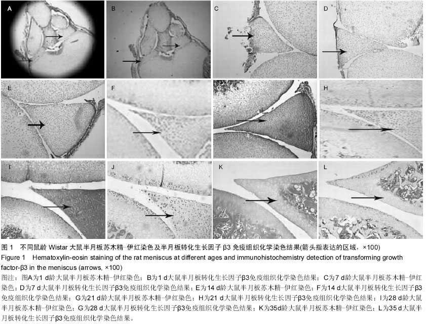
| [1] LaPrade MD, LaPrade CM, Hamming MG, et al. Intramedullary Tibial Nailing Reduces the Attachment Area and Ultimate Load of the Anterior Medial Meniscal Root: A Potential Explanation for Anterior Knee Pain in Female Patients and Smaller Patients. Am J Sports Med.2015; 43(7):1670-1675.
[2] Walker PS, Arno S, Bell C, et al.Function of the medial meniscus in force transmission and stability. J Biomech.2015; 48(8):1383-1388.
[3] McNulty AL, Guilak F.Mechanobiology of the meniscus. J Biomech.2015; 48(8):1469-1478.
[4] Griffin JW, Hadeed MM, Werner BC, et al. Platelet-rich plasma in meniscal repair: does augmentation improve surgical outcomes? Clin Orthop Relat Res.2015; 473(5):1665-1672.
[5] Beaufils P, Becker R, Verdonk R, et al.Focusing on results after meniscus surgery. Knee Surg Sports Traumatol Arthrosc.2015; 23(1):3-7.
[6] Plymale M, Fleisig GS, Kocaj SM, et al. Visualization and reduction of a mensical capsular junction tear in the knee: an arthroscopic surgical technique. Am J Orthop (Belle Mead NJ). 2014; 43(11):498-500.
[7] Khan M, Evaniew N, Bedi A, et al. Arthroscopic surgery for degenerative tears of the meniscus: a systematic review and meta-analysis.CMAJ. 2014; 186 (14):1057-1064.
[8] Grassi A, Zaffagnini S, Marcheggiani Muccioli GM, et al. Clinical outcomes and complications of a collagen meniscus implant: a systematic review. Int Orthop. 2014; 38(9):1945- 1953.
[9] Rubinstein RA Jr, DeHaan A, Baldwin JL.Posterior medial meniscus detachment: a unique type of medial meniscal tear. J Knee Surg.2009; 22(4):339-345.
[10] Samitier G, Alentorn-Geli E, Taylor DC,et al.Meniscal allograft transplantation. Part 2: systematic review of transplant timing, outcomes, return to competition, associated procedures, and prevention of osteoarthritis. Knee Surg Sports Traumatol Arthrosc.2015; 23(1):323-333.
[11] Roos EM, Ostenberg A, Roos H, et al. Long-term outcome of meniscectomy: symptoms, function, and performance tests in patients with or without radiographic osteoarthritis compared to matched controls. Osteoarthritis Cartilage. 2001;9(4): 316-324.
[12] Kobayashi K1, Amiel M, Harwood FL, et al.The long-term effects of hyaluronan during development of osteoarthritis following partial meniscectomy in a rabbit model. Osteoarthritis Cartilage.2000; 8(5):359-365.
[13] Logan M, Watts M, Owen J, et al. Meniscal repair in the elite athlete: results of 45 repairs with a minimum 5-year follow-up. Am J Sports Med.2009; 37(6):1131-1134.
[14] Moore-Olufemi SD, Olsen AB, Hook-Dufresne DM, et al. Transforming growth factor-Beta 3 alters intestinal smooth muscle function: implications for gastroschisis-related intestinal dysfunction. Dig Dis Sci. 2015; 60(5):1206-1214.
[15] Jin JZ, Warner DR, Lu Q, et al. Deciphering TGF-β3 function in medial edge epithelium specification and fusion during mouse secondary palate development. Dev Dyn.2014; 243(12):1536-1543.
[16] Walshe TE, dela Paz NG, D'Amore PA. The role of shear-induced transforming growth factor-β signaling in the endothelium. Arterioscler Thromb Vasc Biol. 2013; 33(11): 2608-2617
[17] Ye P. Modulation of epithelial tight junctions by TGF-beta 3 in cultured oral epithelial cells. Aust Dent J.2012;57(1):11-17.
[18] Harrison CA, Al-Musawi SL, Walton KL. Prodomains regulate the synthesis, extracellular localisation and activity of TGF-β superfamily ligands. Growth Factors. 2011; 29(5):174-186.
[19] Nakamura S, Kawai T, Kamakura T, et al. TGF-beta3 is expressed in taste buds and inhibits proliferation of primary cultured taste epithelial cells. In Vitro Cell Dev Biol Anim.2010; 46(1):36-44.
[20] Adams DH, Ruzehaji N, Strudwick XL, et al. Attenuation of Flightless I, an actin-remodelling protein, improves burn injury repair via modulation of transforming growth factor (TGF)-beta1 and TGF-beta3. Br J Dermatol.2009; 161(2): 326-336.
[21] Steffl M, Schweiger M, Amselgruber WM. Expression of transforming growth factor-beta3 (TGF-beta3) in the porcine ovary during the oestrus cycle. Histol Histopathol.2008; 23(6):665-671.
[22] Mercado-Pimentel ME, Runyan RB. Cells Tissues Organs. Multiple transforming growth factor-beta isoforms and receptors function during epithelial-mesenchymal cell transformation in the embryonic heart.2007;185(1-3): 146-156.
[23] Risbud MV, Di Martino A, Guttapalli A, et al.Toward an optimum system for intervertebral disc organ culture: TGF-beta 3 enhances nucleus pulposus and anulus fibrosus survival and function through modulation of TGF-beta-R expression and ERK signaling. Spine (Phila Pa 1976).2006; 31(8):884-890.
[24] Arnoczky SP, Warren RF, Spivak JM. Meniscal repair using an exogenous fibrin clot. An experimental study in dogs. J Bone Joint Surg Am.1988; 70(8):1209-1217.
[25] Nakata K, Shino K, Hamada M, et al. Human meniscus cell: characterization of the primary culture and use for tissue engineering. Clin Orthop Relat Res. 2001; (391 Suppl): S208-218.
[26] Goldring MB, Tsuchimochi K, Ijiri K. The control of chondrogenesis.J Cell Biochem.2006;97(1):33-44.
[27] Innocenti M, Ceruso M, Manfrini M, et al. Free vascularized growth-plate transfer after bone tumor resection in children.J Reconstr Microsurg. 1998;14(2):137-143.
[28] Parekh T, Saxena B, Reibman J,et al. Neutrophil chemotaxis in response to TGF-beta isoforms (TGF-beta 1, TGF-beta 2, TGF-beta 3) is mediated by fibronectin. J Immunol.1994; 152(5):2456-2466.
[29] Lu SL, Reh D, Li AG,et al. Overexpression of transforming growth factor beta1 in head and neck epithelia results in inflammation, angiogenesis, and epithelial hyperproliferation. Cancer Res. 2004; 64(13):4405-4410.
[30] Cui X, Zeni F, Vodovitz Y,et al. TGF-beta1 increases microbial clearance but worsens lung injury during Escherichia coli pneumonia in rats. Cytokine. 2003; 24(4):115-127. |