| [1] 陆声,徐永清,张元智,等.计算机辅助导航模板在下颈椎椎弓根定位中的临床应用[J].中华骨科杂志,2008,28(12):1002-1007.
[2] 胡勇,马维虎,徐荣明,等.组合枢锥椎板螺钉固定技术治疗颈椎损伤的临床研究[J].中华骨科杂志,2009,25(3):218-222.
[3] Rampersaud YR, Simon DA, Foley KT. Accuracy requirements for image-guided spinal pedicle screw placement. Spine.2001;26(4):352-359.
[4] League D.Interactive, image-guided, stereotactic neurosurgery systems.AORNJ.1995;61(2):360-370.
[5] 杨永宏,郑杰.计算机辅助导航系统及其骨科应用[J].中华创伤骨科杂志,2005,7(7):614-616.
[6] 王军强,孙磊,王满宜.计算机辅助骨科手术的应用和进展[J].中华创伤骨科杂志,2004,6(1):110-114.
[7] 段彦静,孙文磊.逆向工程和快速成型技术及医学应用[J].医药卫生装备,2006,27(10):31-34.
[8] Wang G, Li J, Khadka A. et al. CAD/CAM and rapid prototyped titanium for reconstruction of ramus defect and condylar fracture caused by mandibular reduction. Oral Surg Oral Med oral Pathol Oral Radiol.2012;113(3):356-361.
[9] Levine JP,Patel A,Saadeh PB,et al.Computer-aided design manufacturing in craniomaxillofacial surgery: the new state of the art. J Craniofac Surg. 2012;23(1):288-293.
[10] Harvie P, Chesser TJ, Ward AJ. The Bristol regional pelvic and acetabular fracture service: workload implications of managing the polytraumatised patient. Injury. 2008; 39(8): 839-843.
[11] Whitmarsh T, Fritscher KD, Humbert L,et al. A statistical model of shape and bone mineral density distribution of the proximal femur for fracture risk assessment. Med Image Comput Comput Assist Interv. 2011;14(Pt 2):393-400.
[12] Luo Y, Ferdous Z, Leslie WD. A preliminary dual-energy X-ray absorptiometry-based finite element model for assessing osteoporotic hip fracture risk.Proc Inst Mech Engh.2011; 225(12):1188-1195.
[13] Starker M, Bischof F, Lindenfeld T. Total hip arthroplasty with shortening subtrochanteric osteotomy and custom-made prosthesis in Crowe type IV developmental dysplasia. Z Orthop Unfall. 2011;149(5):518-525.
[14] Nelson JD, Mclff TE, Moodie PG, et al Biomehchanical stability of intramedullay technique for fixation of joint depressed calcaneus fracture.Foot Anke Int. 2010;31(3): 229-235.
[15] Kamalian S, Hirsch AE, Growney ML, et al. CT guided percutaneous calcaneoplasty: a case of metastatic intra-articular calcaneus fracrure. J Neurointerv surg. 2009;1(2):186-188.
[16] Shah M. Auricular prosthesis fabrication using computer-aided design and rapid prototyping technologies. J Prosthet Orthot Int.2013;10(8):1-3.
[17] Tong K, Zhang Y, Zhang S, et al. Application of computer-aided osteotomy template design in treatment of developmental dysplasia of the hip with steel osteotomy. Nan Fang Yi Ke Da Xue Xue Bao. 2013;33(6):906-909.
[18] Faur C, Crainic N,Sticlaru C,et al. Rapid prototyping technique in the preoperative planning for total hip arthroplasty with custom femoral components. J Wien Klin Wochenschr.2013;125(5-6):144-149.
[19] Sun J, Zhang FQ. The application of rapid prototyping in prosthodontics. J Prosthodont. 2012;21(8):641-644.
[20] 林锦乐,林壹冰,黄春育,等.计算机辅助复杂骨盆骨折手术治疗与疗效观察[J].长江大学学报,2011,8(1):146-148.
[21] 涂强,丁焕文,曹露,等.计算机辅助技术在复杂骨盆骨折诊断与治疗方面的应用[J].临床骨科杂志,2011,14(2):204-206.
[22] 胡学峰,洪建明,刘敏等.快速成型技术在骨盆髋臼骨折治疗中的应用研究[J].江西医学院学报,2007,47(5):56-58.
[23] Zheng Z, Zhang Y, Hou Z, et al. The application of a computer-assisted thermoplastic membrane navigation system in screw fixation of the sacroiliac joint--a clinical study. Injury. 2012;43(4):495-499.
[24] Beldame J, Boisrenoult P, Beaufils P. Pin track induced fractures around computer-assisted TKA. Orthop Traumatol Surg Res. 2010;96(3):249-255.
[25] Wysocki RW, Sheinkop MB, Virkus WW, et al. Femoral fracture through a previous pin site after computer-assisted total knee arthroplasty. J Arthroplasty. 2008;23(3):462-465.
[26] Mahomed NN, Arndt DC,McGrory BJ, et al. The Harris hip score: comparison of patient self-report with surgeon assessment.J Arthroplasty.2001;16(5):575-580.
[27] Bagaria V, Deshpande S, Rasalkar DD, et al. Use of rapid prototyping and three-dimensional reconstruction modeling in the management of complex fractures. Eur J Radiol. 2011; 80(3):814-820.
[28] Shen F, Chen B, Guo Q, et al. Augmented reality patient-specific plate design for pelvic and acetabular fracture surgery. J Int J Comput Assist Radiol Surg. 2013;8(2): 169-179.
[29] Stoker NG, Mankovich NJ, Valentino D. Stereolithographic models for surgical planning: preliminary report. J Oral Maxillofac Surg. 1992;50(5):466-471.
[30] Brie J, Chartier T, Chaput C, et al. A new custom made bioceramic implant for the repair of large and complex craniofacial bone defects. J Craniomaxillofac Surg. 2013; 41(5):403-407. |
.jpg)
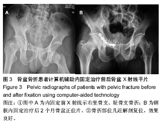
.jpg)
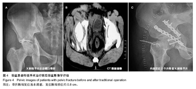
.jpg)