| [1]Toshihiro, Ishijima T, Hashikawa Y, et al. Osteoporosis and reduction of residual ridge in edentulous patients. J Prosthet Dent. 1993;69(1):49.
[2]Bolscher MT, Netelebos JC, Barto R, et al. Estrogen regulation of intestinal calcium absorption in the intact ovariectomized adult rat. J Bone Miner Res. 1999;14: 1197-1202.
[3]Ozmen B, Kirmaz C, Aydin K, et al. Influence of the selective ostrogen receptor modulator on IL-6,TNF-alpha,TGF-beta1 and bone turnover markers in the treatment of postmenopausal osteoporosis. Eur Cytekine Netw. 2007; 18(3):148-153.
[4]李红祥.去势大鼠骨质疏松模型的研究[J]. 现代预防医学,1992, 19(1):5.
[5]王敏,袁绍云. 全身骨密度,下颌骨密度与无牙颌牙槽骨骨吸收关系的初步研究[J].现代口腔医学杂志,1991,8(3):174-176.
[6]朱晓滨,于世凤,史凤芹. 骨质疏松症患者下颌骨骨密度的分析研究[J].现代口腔医学杂志,1996,10(2):78-80.
[7]Wende JW, Sara G, Maurizio G, et al. The role of osteopenia in oral bone loss and periodontal disease. J Periodotol. 1996; 67(105):1076.
[8]杜莉,胡国瑜.大鼠雌激素低下时下颌骨和股骨同位素测定的比较[J].华西口腔医学杂志,1995,13(3):2044.
[9]Shimizu M, Sasaki T, Ishihara A, et al. Bone wound healing after maxillary molar extraction in ovariectomized aged rats. J Electron Microsc. 1998;47(5):517-526.
[10]Tanaka S, Shimizu M, Debari K, et al. Acute effects of ovariectomy on wound healing of alveolar bone after maxillary molar extraction in aged rats. Anat Rec. 2002;262:203-212.
[11]Lim SK, Won YJ, Lee HC, et al. A PCR analysis of ER and ER mRNA abundance in bats and the effect. of ovariectomy. J Bone Miner Res, 1999,14: 1189-1196.
[12]Bolscher MT, Netelebos JC, Barto R, et al. Estrogen regulation of intestinal calcium absorption in the intact and ovariectomized adult rat. J Bone Miner Res, 1999,14: 1197-1202.
[13]Suda T, Nakamara I, Jimi E, et al. Regulation of osteoclast function. J Bone Miner Res. 1997;12: 869-879.
[14]Frost HU. On the estrogen-bone relationship and postmenopausal bone loss: a new model. J Bone Miner Res. 1999;14: 1473-1477.
[15]Ishikawa H, Tanaka H, Iwato K, et al. Effect of glucocorticoids on the biologic activities of myeloma cells: inhibition of interleukin beta osteoclast activating factor-induced bone resorption. Blood. 1990;75:175.
[16]Pfeilschifter J, Chenu C, Bird A, et al. Interleukin-1 and tumorencrosis factor stimulate the formation of human osteoclastlike cells in vitro. J Bone Miner Res.1989;4:11310.
[17]Shen V, Kohler G, Jeffrey J. Human osteoblast in vitro secrete tissue inhibitor of metallo proteinase and gelatinase but not interstitial collagen as major cellular production. J Clin Invest. 1989;84(2):686-694.
[18]Robert B, Kimble JL, Vannice DC, et al. Interleukin-1 receptor antagonist decrease bone loss and resorption in ovariectomized rats. J Clin Invest. 1994;93:1959-1967.
[19]Ellies LG, Aubin JE. Temporal sequence of Interleukin-1-mediated stimulation and inhibition of bone formation by isolated fetal rat calvaria cell in vetro. Cytokine. 1990;2:43017.
[20]李潇, 丁寅, 金作霖,等. 雌激素对骨质疏松大鼠牙槽骨改建及IL-1表达的影响[J].牙体牙髓牙周病学杂志,2000,10(1):16-18. |
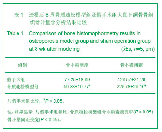
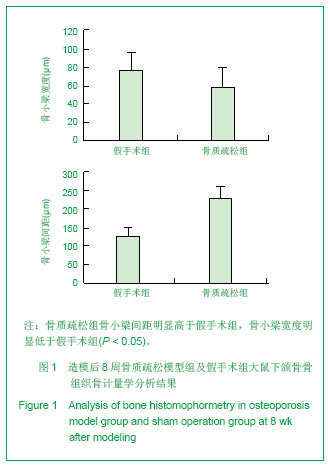

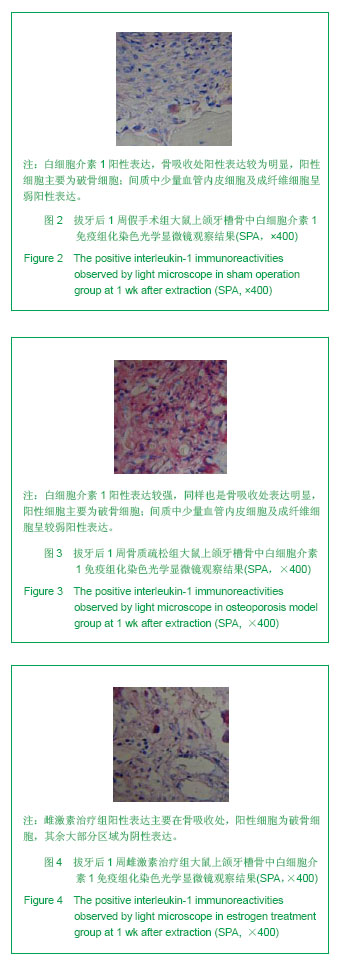


.jpg)