| [1]Küpeli E, Eyübo?lu FÖ, Haberal M.Pulmonary infections in transplant recipients. Curr Opin Pulm Med.2012;18(3): 202-212.
[2]Kotloff RM, Ahya VN, Crawford SW.Pulmonary complications of solid organ and hematopoietic stem cell transplantation. Am J Respir Crit Care Med. 2004; 170(1):22-48.
[3]Batista MV, Costa SF, Shikanai-Yasuda MA, et al. Current treatment options for invasive aspergillosis.Drugs Today (Barc). 2013;49(3):213-226.
[4]Beigelman-Aubry C, Godet C, Caumes E. Lung infections: The radiologist's perspective.Diagn Interv Imaging. 2012; 93(6):431-440.
[5]Kousha M, Tadi R, Soubani AO.Pulmonary aspergillosis: a clinical review.Eur Respir Rev. 2011;20(121):156-1574.
[6]Wichmann D, Kluge S.Invasive pulmonary aspergillosis in the ICU: an emerging disease? Intensive Care Med. 2013 Jan 22.
[7]Smith JA, Kauffman CA.Pulmonary fungal infections. Respirology. 2012;17(6):913-926.
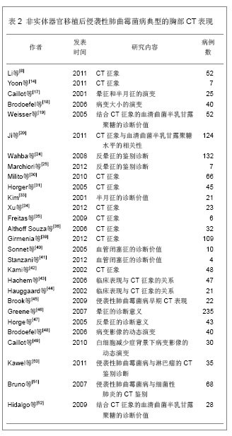
[8]Li XS, Zhu HX, Fan HX, et al.Pulmonary fungal infections after bone marrow transplantation: the value of high-resolution computed tomography in predicting their etiology.Chin Med J (Engl). 2011;124(20):3249-3254.
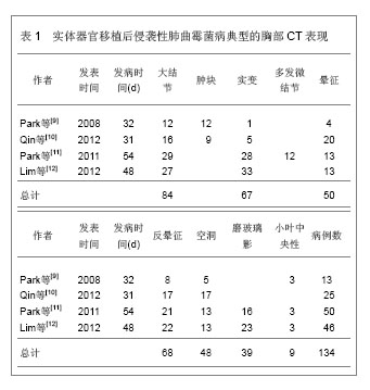
[9]Park YS, Seo JB, Lee YK, et al.Radiological and clinical findings of pulmonary aspergillosis following solid organ transplant. Clin Radiol. 2008;63(6):673-680.
[10]Qin J, Fang Y, Dong Y, et al.Radiological and clinical findings of 25 patients with invasive pulmonary aspergillosis: retrospective analysis of 2150 liver transplantation cases.Br J Radiol. 2012;85(1016):e429-435.
[11]Park SY, Lim C, Lee SO, et al.Computed tomography findings in invasive pulmonary aspergillosis in non-neutropenic transplant recipients and neutropenic patients, and their prognostic value. J Infect. 2011;63(6):447-456.
[12]Lim C, Seo JB, Park SY, et al. Analysis of initial and follo w-up CT findings in patients with invasive pulmonary aspergillosis after solid organ transplantation. Clin Radiol 2012;67(12): 1179-1186.
[13]Greene R.The radiological spectrum of pulmonary aspergillosis. Med Mycol. 2005;43 Suppl 1:S147-154.
[14]Yoon SH, Park CM, Goo JM. Pulmonary aspergillosis in immunocompetent patients without air-meniscus sign and underlying lung disease: CT findings and histopathologic features.Acta Radiol. 2011;52(7):756-761.
[15]Parrón M, Torres I, Pardo M, et al. The halo sign in computed tomography images: differential diagnosis and correlation with pathology findings. Arch Bronconeumol. 2008;44(7): 386-392.
[16]Lee YR, Choi YW, Lee KJ, et al.CT halo sign: the spectrum of pulmonary diseases. Br J Radiol. 2005;78(933):862- 865.
[17]Caillot D, Couaillier JF, Bernard A, et al. Increasing volume and changing characteristics of invasive pulmonary aspergillosis on sequential thoracic computed tomography scans in patients with neutropenia. J Clin Oncol 2001;19(1): 253-259.
[18]Brodoefel H, Vogel M, Hebart H, et al. Long-term CT follow-up in 40 non-HIV immunocompromised patients with invasive pulmonary aspergillosis: kinetics of CT morphology and correlation with clinical findings and outcome. AJR Am J Roentgenol 2006;187(2):404-413.
[19]Weisser M, Rausch C, Droll A, et al. Galactomannan does not precede major signs on a pulmonary computerized tomographic scan suggestive of incasive aspergillosis in patients with hematological malignancies. Clin Infect Dis. 2005;41(8):1143-1149.
[20]Ji Y, Xu LP, Liu DH, et al.Positive results of serum galactomannan assays and pulmonary computed tomography predict the higher response rate of empirical antifungal therapy in patients undergoing allogeneic hematopoietic stem cell transplantation.Biol Blood Marrow Transplant. 2011;17(5): 759-764.
[21] Park SY, Kim SH, Choi SH ,et al.Clinical and radiological features of invasive pulmonary aspergillosis in transplant recipients and neutropenic patients. Transpl Infect Dis. 2010; 12(4):309-315.
[22]Marchiori E, Zanetti G, Hochhegger B, et al.Reversed halo sign on computed tomography: state-of-the-art review. Lung. 2012;190(4):389-394.
[23]Georgiadou SP, Sipsas NV, Marom EM, et al.The Diagnostic Value of Halo and Reversed Halo Signs for Invasive Mold Infections in Compromised Hosts Clin Infect Dis. 2011;52(9): 1144-1155.
[24]Wahba H, Truong MT, Lei X, et al.Reversed halo sign in invasive pulmonary fungal infections. Clin Infect Dis. 2008; 46(11):1733-1737.
[25]Marchiori E, Zanetti G, Escuissato DL, et al.Reversed halo sign: high-resolution CT scan findings in 79 patients. Chest. 2012;141(5):1260-1266.
[26]Godoy MC, Viswanathan C, Marchiori E, et al. The reversed halo sign: update and differential diagnosis.Br J Radiol. 2012; 85(1017):1226-1235.
[27]Marchiori E, Zanetti G, Hochhegger B, et al.Reversed halo sign in active pulmonary tuberculosis: criteria for differentiation from cryptogenic organizing pneumonia.Lung. 2012;190(4):389-394.
[28]Cottin V, Cordier JF.Cryptogenic organizing pneumonia.Semin Respir Crit Care Med. 2012;33(5):462-475.
[29]Marchiori E, Zanetti G, Irion KL,Reversed halo sign on computed tomography: state-of-the-art review.AJR Am J Roentgenol. 2011;197(6):1324-1327.
[30]Milito MA, Kontoyiannis DP, Lewis RE, et al. Influence of host immunosuppression on CT findings in invasive pulmonary aspergillosis. 2010;48(6):817-823.
[31]Horger M, Hebart H, Einsele H, et al.Initial CT manifestations of invasive pulmonary aspergillosis in 45 non-HIV immunocompromised patients: association with patient outcome?Eur J Radiol. 2005;55(3):437-44.
[32]Fred HL, Gardiner CL.The air crescent sign: causes and characteristics. Tex Heart Inst J. 2009;36(3):264-265.
[33].Tonelli AR, Khalife WT, Cao M. Spherules, hyphae, and air-crescent sign.Am J Med Sci. 2008;335(6):504-506.
[34]Xu SC, Qiu LH, Liu WY, et al.Initial computed tomography findings of invasive pulmonary aspergillosis in non-hematological patients.Chin Med J (Engl). 2012; 125(17): 2979-2985.
[35]Freitas DB, Piovesan AC, Szarf G, et al.Outbreak of invasive pulmonary aspergillosis among patients hospitalized in a bone marrow transplant ward: tomographic findings. J Bras Pneumol. 2009;35(9):931-936.
[36]Althoff Souza C, Müller NL, Marchiori E, et al.Pulmonary invasive aspergillosis and candidiasis in immunocompromised patients: a comparative study of the high-resolution CT findings. J Thorac Imaging. 2006;21(3):184-189.
[37]Battista G, Sassi C, Zompatori M, et al.Ground-glass opacity: interpretation of high resolution CT findings. Radiol Med. 2003; 106(5-6):425-444.
[38]Diederich S. High resolution computed tomography of the lungs: ground glass opacity and its differential diagnosis. Radiologe. 2010;50(12):1141-1152.
[39]Girmenia C, Guerrisi P, Frustaci AM, et al.New category of probable invasive pulmonary aspergillosis in haematological patients.Clin Microbiol Infect. 2012;18(10):990-996.
[40] Sonnet S, Buitrago-Tellez CH, Tamm M, et al. Direct detection of angioinvasive pulmonary aspergillosis in immunosuppressed patients: preliminary results with high-resolution 16-MDCT angiography. AJR Am J Roentgenol. 2005;184(3):746-751.
[41] Stanzani M, Battista G, Sassi C, et al.Computed tomographic pulmonary angiography for diagnosis of invasive mold diseases in patients with hematological malignancies. Clin Infect Dis. 2012;54(5):610-616.
[42]Kami M, Kishi Y, Hamaki T. The value of the chest computed tomography halo sign in the diagnosis of invasive pulmonary aspergillosis. An autopsy-based retrospective study of 48 patients. Mycoses. 2002;45(8):287-294.
[43]Hachem R, Sumoza D, Hanna H. Clinical and radiologic predictors of invasive pulmonary aspergillosis in cancer patients: should the European Organization for Research and Treatment of Cancer/Mycosis Study Group (EORTC/MSG) criteria be revised? Cancer. 2006;106(7):1581-1586.
[44]Hauggaard A, Ellis M, Ekelund L. Early chest radiography and CT in the diagnosis, management and outcome of invasive pulmonary aspergillosis.Acta Radiol. 2002;43(3):292-298.
[45]Brook O, Guralnik L, Hardak E. Radiological findings of early invasive pulmonary aspergillosis in immune-compromised patients.Hematol Oncol. 2009;27(2):102-106.
[46]Greene RE, Schlamm HT, Oestmann JW. Imaging findings in acute invasive pulmonary aspergillosis: clinical significance of the halo sign. Clin Infect Dis. 2007;44(3):373-379.
[47]Horger M, Einsele H, Schumacher U. Invasive pulmonary aspergillosis: frequency and meaning of the "hypodense sign" on unenhanced CT. Br J Radiol. 2005;78(932):697-703.
[48]Brodoefel H, Vogel M, Hebart H. Long-term CT follow-up in 40 non-HIV immunocompromised patients with invasive pulmonary aspergillosis: kinetics of CT morphology and correlation with clinical findings and outcome. AJR Am J Roentgenol. 2006;187(2):404-413.
[49]Caillot D, Latrabe V, Thiébaut A. Computer tomography in pulmonary invasive aspergillosis in hematological patients with neutropenia: an useful tool for diagnosis and assessment of outcome in clinical trials.Eur J Radiol. 2010;74(3): e172-175.
[50]Kawel N, Schorer GM, Desbiolles. Discrimination between invasive pulmonary aspergillosis and pulmonary lymphoma using CT.Eur J Radiol. 2011;77(3):417-425.
[51]Bruno C, Minniti S, Vassanelli A. Comparison of CT features of Aspergillus and bacterial pneumonia in severely neutropenic patients. J Thorac Imaging. 2007;22(2):160-165.
[52]Hidalgo A, Parody R, Martino R. Correlation between high-resolution computed tomography and galactomannan antigenemia in adult hematologic patients at risk for invasive aspergillosis. Eur J Radiol. 2009;71(1):55-60.
[53]Kwon JC, Kim SH, Park SH, et al. Prognosis of invasive pulmonary aspergillosis in patients with hematologic diseases in Korea.Tuberc Respir Dis (Seoul). 2012;72(3):284-292.
[54]Kurosawa M, Yonezumi M, Hashino S, et al. Epidemiology and treatment outcome of invasive fungal infections in patients with hematological malignancies.Int J Hematol. 2012;96(6):748-757.
[55]Herbrecht R, Roedlich MN. Earlier diagnosis of angioinvasive pulmonary mold disease: is computed tomography pulmonary angiography a new step? Clin Infect Dis. 2012;54(5):610-616. |