| [1] Hsu JY, Bourguignon LY, Adams CM, et al. Matrix metalloproteinase-9 facilitates glial scar formation in the injured spinal cord.J Neurosic.2008;28(50): 13467-13477.
[2] Su Z, Yuan Y, Chen J, et al. Reactive astrocytes inhibit the survival and differentiation of oligodendrocyte precursor cells by secreted TNF-alpha.J Neurotrauma.2011;28(6): 1089-100.
[3] Colangelo AM. Targeting reactive astrogliosis by novel biotechnological strategies. Biotechnol Adv.2011;6(5): 5.
[4] Silver J, Miller JH. Regeneration beyond the glial scar.Nat Rev Neurosci. 2004;5(2):146-156.
[5] Vos PE,Jacobs B,Andriessen TM,et al.GFAP and S100B are biomarkers of traumatic brain injury: an observational cohort study. Neurology. 2010;75(20):1786-1793.
[6] Dixon KJ, Munro KM, Boyd AW, et al. Partial change in EphA4 knockout mouse phenotype: loss of diminished GFAP upregulation following spinal cord injury.Neurosci Lett.2012; 525(1):66-71.
[7] 田代实,王伟,徐运兰,等.细胞周期素依赖性激酶抑制剂olomoucine对大鼠脊髓损伤后轴突再生微环境的影响及其意义[J].中华医学杂志,2006,86(13):901-905.
[8] 王春婷,贺诗华,游思维,等.自组装肽包裹嗅神经鞘细胞抑制脊髓损伤后胶质瘢痕形成并促进轴突再生[J].解剖学研究, 2009, 31(6):401-405.
[9] Yamamoto M, Raisman G, Li D, et al. Transplanted olfactory mucosal cells restore paw reaching function without regeneration of severed corticospinal tract fibres across the lesion.Brain Res.2009;1303:26-31.
[10] Ribotta MG,Menet V,Privat A.Glial scar and axonal regeneration in the CNS : lessons from GFAP and vimentin t ransgenic mice. Acta Neurochir Suppl. 2004;89:87292.
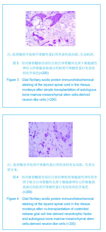
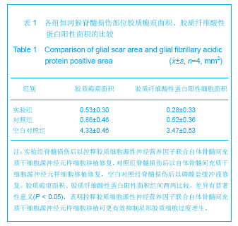
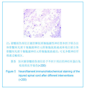
[11] 刘晓刚,邓宇斌,刘祖国,等.神经元样细胞与控释神经营养因子联合移植对脊髓损伤猴后索结构修复和功能恢复的作用[J].中国临床康复, 2005, 9(26):96-99.
[12] 刘晓刚,邓宇斌,蔡辉,等. 控释胶质细胞源性神经营养因子对骨髓间质干细胞源性神经元样细胞移植修复猴脊髓轴突的协同作用[J].中国组织工程研究与临床康复, 2007, 11(11): 2053-2056.
[13] 刘晓刚,邓宇斌,蔡辉,等.诱发电位在GDNF与MSCs联合移植法治疗猴脊髓损伤评价中的应用[J].中国康复医学杂志,2009, 24(6):509-511.
[14] 刘晓刚, 邓宇斌, 蔡辉, 等.控释胶质细胞源性神经营养因子与骨髓间充质干细胞源性神经元样细胞移植对猴损伤脊髓髓鞘的修复[J].中国组织工程研究与临床康复, 2009, 13(32): 6219-6222.
[15] 刘晓刚,邓宇斌,刘祖国,等. 骨髓间质干细胞源性早期神经元与控释神经营养因子移植治疗猴脊髓损伤的研究[J].中华神经外科杂志, 2006,22(10):599-601.
[16] Kamei N, Kwon SM, Ishikawa M, et al. Endothelial progenitor cells promote astrogliosis following spinal cord injury through Jagged1-dependent Notch signaling.J Neurotrauma.2012; 29(9):1758-1769.
[17] Sofroniew MV. Molecular dissection of reactive astrogliosis and glial scar formation. Trends Neurosci.2009;32(12): 638-647.
[18] Liu C, Wu ZZ, Shu CL, et al. Experimental investigation of HGF inhibiting glial scar in vitro.Cell Mol Neurobiol.2011;31(2): 259-268.
[19] Leal-Filho MB. Spinal cord injury: From inflammation to glial scar.Surg Neurol Int. 2011; 2: 112.
[20] 陈安,张建伟,王慧,等. 建立中枢神经再生大鼠红核脊髓束横断模型的研究[J]. 湖南中医药大学学报,2006,26(6):9210.
[21] Yamaoka G, Morino T, Morizane K,et al. p38 mitogen-activated protein kinase inhibitor reduces neurocan production in cultured spinal cord astrocytes.Neuroreport. 2012;23(9):546-550.
[22] Tsai HH, Li H, Fuentealba LC, et al. Regional astrocyte allocation regulates CNS synaptogenesis and repair. Science. 2012;337(6092):358-362.
[23] Halassa MM. Synaptic islands defined by the territory of a single astrocyte.J Neurosci.2007;27(24): 6473-6477.
[24] Laywell ED, Rakic P, Kukekov VG, et al. Identification of a multipotent astrocytic stem cell in the immature and adult mouse brain.Proc Natl Acad Sci USA.2000;97(25): 13883-13888.
[25] Schwab JM, Brechtel K, Mueller CA, et al. Experimental strategies to promote spinal cord regeneration—an integrative perspective.Prog Neurobiol.2006;78(2): 91-116.
[26] Buss A,Brook GA,Kakulas B,et al.Gradual loss of myelin and formation of an astrocytic scar during Wallerian degeneration in the human spinal cord.Brain.2004;127:34-44.
[27] Anders JJ,Hurlock JA .Transplanted glial scar impedes olfactory bulb reinnervation.Exp Neurol.1996;142(1): 144-150.
[28] Wanner IB, Deik A, Torres M,et al. A new in vitro model of the glial scar inhibits axon growth.Glia.2008;56(15):1691-1709.
[29] Mckeon RJ,Turynec MJ,Buck CR.The Chondroitin Sulfate Proteoglycans Neurocan and Phosphacan Are Expressed by Reactive Astrocytes in the Chronic CNS Glial Scar.J Neurosic. 1999;19(24):10778-10788.
[30] Otsuru S,Hofmann TJ,Olson TS,et al.Improved isolation and expansion of bone marrow mesenchymal stromal cells using a novel marrow filter device. Cytotherapy.2013;15(2): 146-153.
[31] Jacobs SA,Roobrouck VD,Verfaillie CM,et al.Immunological characteristics of human mesenchymal stem cells and multipotent adult progenitor cells.Immunol Cell Biol.2013; 91(1): 32-39.
[32] Fekete N,Rojewski MT,Fürst D,et al. GMP-compliant isolation and large-scale expansion of bone marrow-derived MSC. PLoS One.2012;7(8):e43255.
[33] Kassem M. Mesenchymal stem cells: biological characteristics and potential clinical applications.Cloning Stem Cells.2004;6(4):369-374.
[34] Zhu H,Guo ZK,Jiang XX,et al.A protocol for isolation and culture of mesenchymal stem cells from mouse compact bone. Nat Protoc. 2010;5(3):550-560.
[35] 项鹏, 李树浓. 骨髓间质干细胞研究进展[J].中国病理生理杂志, 2002, 18(11):1438-1442.
[36] Curran JM,Chen R,Hunt JA. Material induced mesenchymal stem cell differentiation.Biomaterials.2010;31(6):1463-1464.
[37] Satija NK, Sharma D, Afrin F,et al. High Throughput Transcriptome Profiling of Lithium Stimulated Human Mesenchymal Stem Cells Reveals Priming towards Osteoblastic Lineage. PLoS One.2013;8(1):e55769.
[38] Durán Alonso MB,Feijoo-Redondo A,Conde de Felipe M,et al.Generation of inner ear sensory cells from bone marrow-derived human mesenchymal stem cells.Regen Med.2012;7(6):769-783.
[39] Egusa H, Kobayashi M, Matsumoto T,et al. Application of cyclic strain for accelerated skeletal myogenic differentiation of mouse bone marrow-derived mesenchymal stromal cells with cell alignment.Tissue Eng Part A.2013;19(5-6):770-782.
[40] Deng Y,Li TQ,Yan YE,et al. Effect of nicotine on chondrogenic differentiation of rat bone marrow mesenchymal stem cells in alginate bead culture.Biomed Mater Eng.2012;22(1-3):81-87.
[41] Ye D,Tanthanuch W,Thumanu K,et al. Discrimination of functional hepatocytes derived from mesenchymal stem cells using FTIR microspectroscopy.Analyst.2012;137(20): 4774-4784.
[42] Grajales L,García J,Geenen DL.Induction of cardiac myogenic lineage development differs between mesenchymal and satellite cells and is accelerated by bone morphogenetic protein-4.J Mol Cell Cardiol.2012;53(3):382-391.
[43] Mantovani C,Terenghi G,Shawcross SG.Isolation of adult stem cells and their differentiation to schwann cells. Methods Mol Biol.2012;916:47-57.
[44] Zhao Y,Xin J,Sun C, et al. Safrole oxide induced neuronal differentiation of rat bone-marrow mesenchymal stem cells by elevating Hsp70.Gene.2012;509(1):85-92.
[45] Wang S,Kan Q,Sun Y,et al. Caveolin-1 regulates neural differentiation of rat bone mesenchymal stem cells into neurons by modulating Notch signaling.Int J Dev Neurosci.2013;31(1):30-35.
[46] Kitada M. Mesenchymal cell populations: development of the induction systems for Schwann cells and neuronal cells and finding the unique stem cell population.Anat Sci Int.2012; 87(1): 24-44.
[47] Wang Y, He W, Bian H,et al. Small molecule induction of neural-like cells from bone marrow-mesenchymal stem cells. J Cell Biochem. 2012;113(5):1527-1536.
[48] Zhang YQ,He LM, Xing B,et al. Neurotrophin-3 gene-modified Schwann cells promote TrkC gene-modified mesenchymal stem cells to differentiate into neuron-like cells in poly(lactic-acid-co-glycolic acid) multiple-channel conduit.Cells Tissues Organs.2012;195(4):313-322.
[49] Seo JH,Cho SR. Neurorestoration induced by mesenchymal stem cells: potential therapeutic mechanisms for clinical trials.Yonsei Med J.2012;53(6):1059-1067.
[50] Sadan O,Shemesh N,Barzilay R,et al.Mesenchymal stem cells induced to secrete neurotrophic factors attenuate quinolinic acid toxicity: a potential therapy for Huntington's disease.Exp Neurol.2012;234(2):417-427.
[51] Shi C. Recent progress toward understanding the physiological function of bone marrow mesenchymal stem cells.Immunology.2012;136(2):133-138.
[52] Arca T,Proffitt J,Genever P. Analysis of human mesenchymal stem cells on a cross-linked collagen-based surgical implant material.Biomed Mater Eng. 2012;22(5):261-276.
[53] Coseo NM,Saldua N,Harrop J. Current use of biologic graft extenders for spinal fusion. J Neurosurg Sci. 2012;56(3): 203-207.
[54] Liu Y, Zhang BA, Song Y, et al. Bone marrow mesenchymal stem cell transplantation for treatment of spinal cord injury: an in vivo magnetic resonance imaging tracking study. Neural Regen Res. 2011;6(13):978-982.
[55] Nakajima H,Uchida K,Guerrero AR,et al. Transplantation of mesenchymal stem cells promotes an alternative pathway of macrophage activation and functional recovery after spinal cord injury.J Neurotrauma.2012;29(8):1614-1625.
[56] Novikova LN,Brohlin M,Kingham PJ,et al. Neuroprotective and growth-promoting effects of bone marrow stromal cells after cervical spinal cord injury in adult rats.Cytotherapy. 2011;13(7):873-887.
[57] Wang D, Zhang JJ. Electrophysiological functional recovery in a rat model of spinal cord hemisection injury following bone marrow-derived mesenchymal stem cell transplantation under hypothermia. Neural Regen Res. 2012;7(10): 749-755.
[58] Alexanian AR,Kwok WM,Pravdic D,et al.Survival of neurally induced mesenchymal cells may determine degree of motor recovery in injured spinal cord rats.Restor Neurol Neurosci. 2010;28(6):761-767.
[59] Chu TH, Wang L, Guo A, et al. GDNF-treated acellular nerve graft promotes motoneuron axon regeneration after implantation into cervical root avulsed spinal cord. Neuropathol Appl Neurobiol.2012;38(7):681-695.
[60] Li L, Lü G, Wang YF, et al. Glial cell-derived neurotrophic factor mRNA expression in a rat model of spinal cord injury following bone marrow stromal cell transplantation. Neural Regen Res 2008;3(10):1056-1059.
[61] Yoo YM, Lee CJ, Kim YJ. Exogenous GDNF increase the migration of the neural stem cells with no protection against kainic acid-induced excitotoxic cell death in rats.Brain Res. 2012;1486:27-38 .
[62] Mahoney MJ,Saltzman WM. Transplantation of brain cells assembled around a programmable synthetic microenvironment.Nat Biotechnol.2001;19(10):934-939.
[63] Bhalala OG, Pan L, Sahni V, et al. microRNA-21 Regulates Astrocytic Response Following Spinal Cord Injury.J Neurosci. 2012;32(50):17935-17947. |

.jpg)