中国组织工程研究 ›› 2013, Vol. 17 ›› Issue (42): 7414-7419.doi: 10.3969/j.issn.2095-4344.2013.42.012

• 材料生物相容性 material biocompatibility • 上一篇    下一篇

磁性壳聚糖微球的生物相容性

王子妤1,董  菊1,张东生2   

  1. 1南京中医药大学基础医学院,江苏省南京市  210023
    2东南大学医学院,江苏省南京市 210009
  • 收稿日期:2013-03-09 修回日期:2013-03-28 出版日期:2013-10-15 发布日期:2013-10-31
  • 作者简介:王子妤☆,女,1979年生,汉族,江苏省无锡市人, 2012年东南大学医学院毕业,讲师,博士,主要从事纳米药物治疗肿瘤方面的研究。 wangziyu717@163.com
  • 基金资助:

    国家自然科学基金资助项目(81201187)*,南京中医药大学基础青年科技创新基金资助项目(10JCQN08)*

Biocompatibility of magnetic chitosan microspheres

Wang Zi-yu1, Dong Ju1, Zhang Dong-sheng2   

  1. 1School of Basic Medical Science, Nanjing University of Chinese Medicine, Nanjing  210023, Jiangsu Province, China
    2School of Medical Science, Southeast University, Nanjing  210009, Jiangsu Province, China
  • Received:2013-03-09 Revised:2013-03-28 Online:2013-10-15 Published:2013-10-31
  • About author:Wang Zi-yu, M.D., Lecturer, School of Basic Medical Science, Nanjing University of Chinese Medicine, Nanjing 210023, Jiangsu Province, China wangziyu717@163.com
  • Supported by:

    the National Natural Science Foundation of China, No. 81201187*; Basic Youth Science and Technology Innovation Fund Project of Nanjing University of Chinese Medicine, No. 10JCQN08*

摘要:

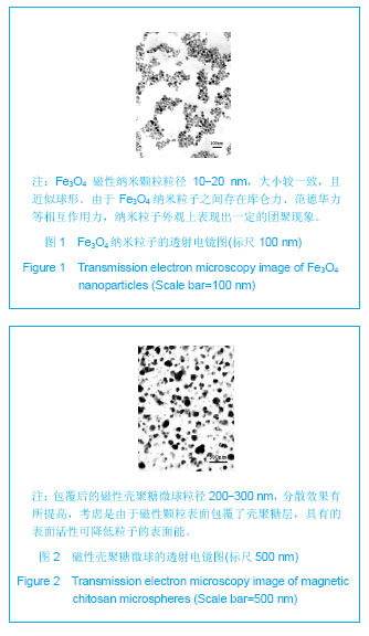
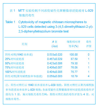
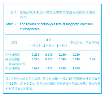
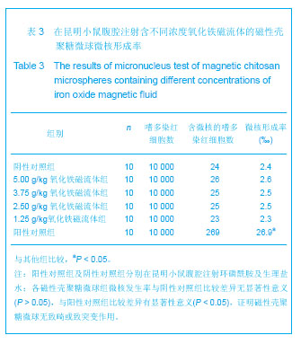
背景:采用壳聚糖对磁性氧化铁纳米颗粒表面进行改性,一方面可改善磁性纳米氧化铁颗粒的团聚性,增加其稳定性,另一方面将来可用于肿瘤热疗及基因治疗。
目的:制备将来可用于肿瘤热疗及基因治疗的磁性壳聚糖微球,评价其生物相容性。
方法:采用改良的化学沉淀法制备磁性氧化铁纳米颗粒。采用超声乳化法将壳聚糖加入到磁性氧化铁纳米颗粒中制备磁性壳聚糖微球,进行以下实验:①MTT实验检测磁性壳聚糖微球浸提液的细胞毒性:分别以1640培养液、聚丙烯酰胺单体溶液、100%,75%,50%,25%的磁性壳聚糖微球浸提液培养L-929细胞。②溶血实验:在兔抗凝血中分别加入磁性壳聚糖微球浸提液、生理盐水及蒸馏水。③微核实验:在昆明小鼠腹腔分别注射含氧化铁磁流体5,3.75,2.5,1.25 g/kg的磁性壳聚糖微球混悬液、环磷酰胺及生理盐水。
结果与结论:磁性壳聚糖微球粒径200-300 nm,分散效果有所提高。不同浓度的磁性壳聚糖微球浸提液对L-929 细胞毒性为1级,属对细胞无毒性范畴。磁性壳聚糖微球浸提液的溶血率为0.69%,小于5%,符合医用材料的溶血实验要求。磁性壳聚糖微球混悬液未导致细胞DNA断裂和非整倍体化,未导致微核产生的遗传毒理作用,材料无致畸或致突变作用,因此磁性壳聚糖微球具有良好的生物相容性。

关键词: 生物材料, 材料生物相容性, 细胞外基质材料, 磁流体, 壳聚糖, 磁性壳聚糖微球, 生物相容性, L-929细胞, 国家自然科学基金

Abstract:

BACKGROUND: Chitosan modification for magnetic iron oxide nanoparticles can improve the agglomeration and stability of magnetic iron oxide nanoparticles, and in addition, it can be used for tumor hyperthermia and gene therapy in the future.
OBJECTIVE: To explore the biocompatibility of magnetic chitosan microspheres which have the potential application in tumor hyperthermia and gene therapy.
METHODS: Magnetic iron oxide nanoparticles were prepared using the modified chemical precipitation. Phacoemulsification method was used to add chitosan into magnetic iron oxide nanoparticles in the preparation of magnetic chitosan microspheres. Then, the following tests were performed. (1) Cytotoxicity test: L-929 cells were cultured in 1640 medium, polyacrylamide monomer solution, 100%, 75%, 50%, 25% of magnetic chitosan microsphere extracts. (2) Hemolysis test: Magnetic chitosan microspheres leaching solution, saline and distilled water were added. (3) Micronucleus test: Kunming mice were intraperitoneally injected with magnetic chitosan microspheres suspension containing 5, 3.75, 2.5, 1.25 g/kg iron oxide magnetic fluid, cyclophosphamide and saline, respectively.
RESULTS AND CONCLUSION: Magnetic chitosan microspheres of 200-300 nm in diameter had an increased dispersion effect. The result of 3-(4,5-dimethylthiazol-2-yl)-2,5-diphenyltetrazolium bromide assay showed that the toxicity of the material on L-929 cell lines belonged to no cytotoxicity. In the hemolysis test, the hemolysis rate of magnetic chitosan microspheres was 0.69% far less than 5%. In the micronucleus test, magnetic chitosan microspheres suspension did not cause DNA fragmentation and aneuploidy, and did not result in genetic toxicological effects produced by micronuclei. The magnetic chitosan microspheres did not appear with cacogenesis and mutagenesis. From the results, magnetic chitosan microspheres are a kind of high biocompatibility material.

Key words: microspheres, chitosan, biocompatible materials, bone matrix

中图分类号: