| [1] Alsberg E, Anderson KW, Albeiruti A,et al.Engineering growing tissues.Proc Natl Acad Sci U S A. 2002;99(19): 12025-12030.
[2] Fishman JM, De Coppi P, Elliott MJ,et al.Airway tissue engineering.Expert Opin Biol Ther. 2011;11(12):1623-1635.
[3] Risbud MV, Sittinger M.Tissue engineering: advances in in vitro cartilage generation.Trends Biotechnol. 2002;20(8): 351-356.
[4] Marcacci M, Filardo G, Kon E.Treatment of cartilage lesions: what works and why. Injury. 2013;44 Suppl 1:S11-15.
[5] Glatz F, Neumeister M, Suchy H,et al. A tissue-engineering technique for vascularized laryngotracheal reconstruction. Arch Otolaryngol Head Neck Surg. 2003;129(2):201-206.
[6] Gilpin DA, Weidenbecher MS, Dennis JE.Scaffold-free tissue-engineered cartilage implants for laryngotracheal reconstruction.Laryngoscope. 2010;120(3):612-617.
[7] 冷晔.聚羟基丁酸己酸酯及其在组织工程中的应用[J].生物医学工程学进展,2011,32(4):213-218.
[8] Chen GQ, Wu Q.The application of polyhydroxyalkanoates as tissue engineering materials.Biomaterials. 2005;26(33): 6565-6578.
[9] 孙安科,胡平,李万同,等.组织工程PHBHH多孔材料喉支架的制备与细胞相容性研究[J].听力学及言语疾病杂志,2011,19(6): 558-561.
[10] Ye C, Hu P, Ma MX,et al.PHB/PHBHHx scaffolds and human adipose-derived stem cells for cartilage tissue engineering. aterials. 2009;30(26):4401-4406.
[11] 孙安科,李万同,孟庆延,等. 带蒂肌筋膜瓣充填与包裹构建喉支架形态组织工程软骨[J].中华耳鼻咽喉头颈外科杂志,2011, 46(12): 1019-1023.
[12] Chesterman PJ, Smith AU. Homotransplantation of articular cartilage and isolated chondrocytes. An experimental study in rabbits. J Bone Joint Surg Br. 1968;50(1):184-197.
[13] Green WT Jr. Articular cartilage repair. Behavior of rabbit chondrocytes during tissue culture and subsequent allografting. Clin Orthop Relat Res. 1977;(124):237-250.
[14] Gardner OF, Archer CW, Alini M,et al. Chondrogenesis of mesenchymal stem cells for cartilage tissue engineering. Histol Histopathol. 2013;28(1):23-42.
[15] Awad HA, Wickham MQ, Leddy HA, et al. Chondrogenic differentiation of adipose-derived adult stem cells in agarose, alginate, and gelatin scaffolds. Biomaterials. 2004;25(16): 3211-3222.
[16] Kramer J, Hegert C, Guan K,et al. Embryonic stem cell-derived chondrogenic differentiation in vitro: activation by BMP-2 and BMP-4.Mech Dev. 2000;92(2):193-205.
[17] Dunham BP, Koch RJ.Basic fibroblast growth factor and insulinlike growth factor I support the growth of human septal chondrocytes in a serum-free environment.Arch Otolaryngol Head Neck Surg. 1998;124(12):1325-1330.
[18] Kim DY, Pyun J, Choi JW,et al. Tissue-engineered allograft tracheal cartilage using fibrin/hyaluronan composite gel and its in vivo implantation.Laryngoscope. 2010;120(1):30-38.
[19] Sterodimas A, de Faria J. Human auricular tissue engineering in an immunocompetent animal model. Aesthet Surg J. 2013; 33(2):283-289.
[20] 孙安科,裴国献,胡平,等.组织工程化喉软骨的构建[J].中华耳鼻咽喉科杂志,2004,39(10):606-611.
[21] 王健,夏万尧,崔磊,等.同种异体组织工程化软骨的免疫学差异[J].中华创伤杂志,2001,17(7):428-430.
[22] 张世浩,朱立新,靳安民,等. 软骨细胞复合胶原/壳聚槲/β-磷酸三钙层状梯度修复体的体外培养[J].中国组织工程研究与临床康复,2008,12(41):8033-8036.
[23] Cohen SB, Meirisch CM, Wilson HA,et al.The use of absorbable co-polymer pads with alginate and cells for articular cartilage repair in rabbits. Biomaterials. 2003;24(15): 2653-2660.
[24] Suwantong O, Waleetorncheepsawat S, Sanchavanakit N,et al. In vitro biocompatibility of electrospun poly(3-hydroxybutyrate) and poly(3-hydroxybutyrate-co-3- hydroxyvalerate) fiber mats. Int J Biol Macromol. 2007;40(3): 217-223.
[25] 刘清宇,王富友,杨柳.关节软骨组织工程支架的研究进展[J].中国修复重建外科杂志,2012,26(10):1247-1250.
[26] Chen GQ, Wu Q.The application of polyhydroxyalkanoates as tissue engineering materials.Biomaterials. 2005;26(33): 6565-6578.
[27] Zhao K, Deng Y, Chun Chen J, et al. Polyhydroxyalkanoate (PHA) scaffolds with good mechanical properties and biocompatibility.Biomaterials. 2003;24(6):1041-1045.
[28] Köse GT, Korkusuz F, Ozkul A,et al.Tissue engineered cartilage on collagen and PHBV matrices.Biomaterials. 2005;26(25):5187-5197.
[29] 王淑芳,郭天瑛,杨超,等. 微生物合成的β-羟基丁酸酯与β-羟基已酸酯共聚物/聚乳酸共混材料(PHBHHx/PLA)的力学性能与生物降解性研究[J].离子交换与吸附,2006,22(1):1-8.
[30] Luo L, Wei X, Chen GQ.Physical properties and biocompatibility of poly(3-hydroxybutyrate-co-3-hydroxyhexanoate) blended with poly(3-hydroxybutyrate-co-4-hydroxybutyrate).J Biomater Sci Polym Ed. 2009;20(11):1537-1553.
[31] 郭霄飞,张永红,杜美丽,等.羟基丁酸与羟基辛酸共聚物一体化骨软骨组织工程支架的制备及性能[J].中国组织工程研究,2012,16(16):2865-2868.
[32] Wang Y, Bian YZ, Wu Q,et al. Evaluation of three-dimensional scaffolds prepared from poly(3-hydroxybutyrate-co-3- hydroxyhexanoate) for growth of allogeneic chondrocytes for cartilage repair in rabbits.Biomaterials. 2008;29(19): 2858-2868.
[33] Deng Y, Lin XS, Zheng Z,et al. Poly(hydroxybutyrate-co- hydroxyhexanoate) promoted production of extracellular matrix of articular cartilage chondrocytes in vitro. Biomaterials. 2003;24(23):4273-4281.
[34] 刘夏君,彭程,肖涛.生物可降解材料构建组织工程软骨的研究进展[J].中国生物工程杂志,2006,26(12):99-106.
[35] Jing Xi, Ling Zhang, Zhenhu An Zheng,et al. Preparation and evaluation of porous poly(3-hydroxybutyrate-co-3- hydroxyhexanoate) hydroxyapatite composite scaffolds.J Biomater Appl. 2008;22(4):293-307.
[36] Cool SM, Kenny B, Wu A, et al. Poly(3-hydroxybutyrate-co-3- hydroxyvalerate) composite biomaterials for bone tissue regeneration: in vitro performance assessed by osteoblast proliferation, osteoclast adhesion and resorption, and macrophage proinflammatory response.J Biomed Mater Res A. 2007;82(3):599-610.
[37] Veleirinho B, Coelho DS, Dias PF, et al. Nanofibrous poly(3-hydroxybutyrate-co-3-hydroxyvalerate)/chitosan scaffolds for skin regeneration. Int J Biol Macromol. 2012; 51(4):343-350.
[38] 高峰,胡平,周建业,等. p(3HB-co-3HH)/PU生物可降解支架材料生物相容性的研究[J].北京生物医学工程,2006,25(3):300-303.
[39] 周栋,胡平,汪钢,等.聚羟基丁酸及其共聚物在体内组织相容性和降解率的实验研究[J].武警医学,2003,14(4):210-212. |
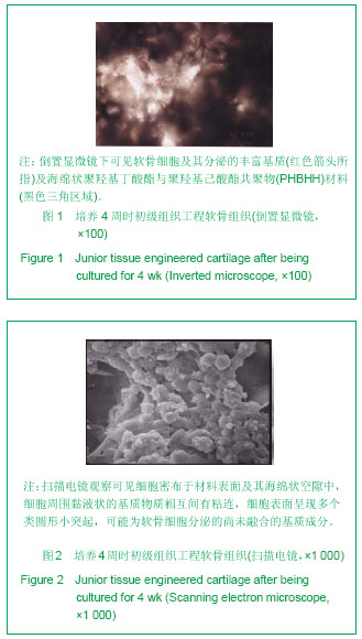
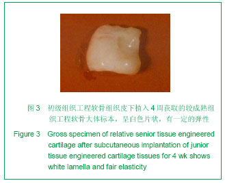
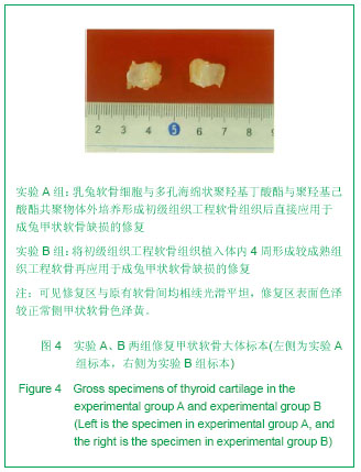
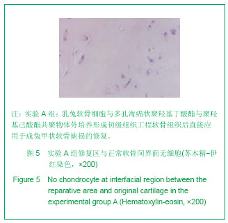
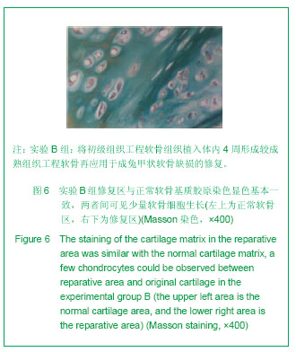
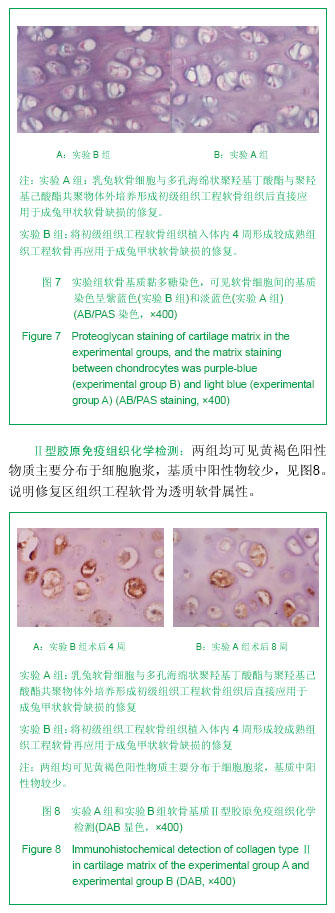
.jpg)