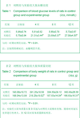
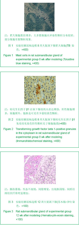
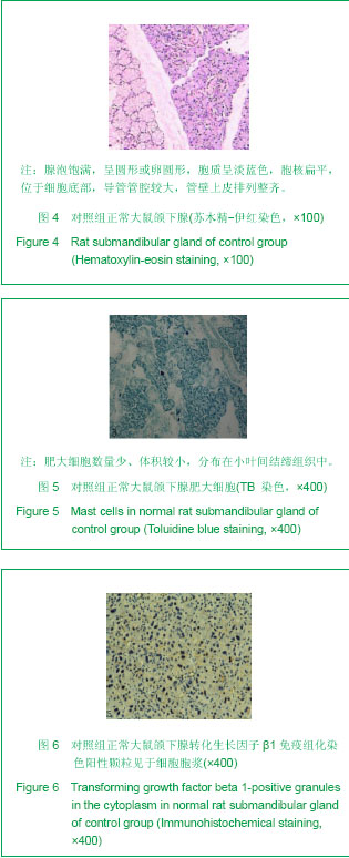
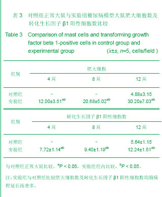
| [1] Atkinson MA, Eisenbarth GS.Type 1 diabetes: new perspectives on disease pathogenesis and treatment. Lancet. 2001;358(9277):221-229.http://www.ncbi.nlm.nih.gov/pubmed?term=.Type%201%20diabetes%3A%20new%20perspectives%20on%20disease%20pathogenesis%20and%20treatment.%20Lancet.[2]Hao JM,Yang ZP. Xiandai Kouqiang Yixue Zazhi. 2001;15(5):373-374.郝京梅,杨宗萍. 2-型糖尿病病人唾液分泌功能与血糖状态间的关系[J].现代口腔医学杂志,2001,15(5):373-374.http://www.cnki.net/KCMS/detail/detail.aspx?QueryID=0&CurRec=1&recid=&filename=XDKY200105028&dbname=cjfd2001&DbCode=CJFQ&urlid=&yx=[3] Gresik EW, Hosoi K, Kurihara K,et al. The rodent granular convoluted tubule cell--an update. Eur J Morphol. 1996;34(3):221-224.http://www.ncbi.nlm.nih.gov/pubmed?term=The%20rodent%20granular%20convaluted%20tubule%20cell-an%20update[4] Amano O, Iseki S. Expression, localization and developmental regulation of insulin-like growth factor I mRNA in rat submandibular gland.Arch Oral Biol. 1993;38(8):671-677.http://www.ncbi.nlm.nih.gov/pubmed/8215990[5] Amano O, Matsumoto K, Nakamura T, et al. Expression and localization of hepatocyte growth factor in rat submandibular gland. Growth Factors. 1994;10(2):145-151.http://www.ncbi.nlm.nih.gov/pubmed/8068352[6] Mogi M, Matsuura S, Suzuki K, et al. Differential expression of transforming growth factor-alpha and epidermal growth factor during postnatal development of rat submandibular gland. Biochem Biophys Res Commun. 1995;217(1):271-277.http://www.ncbi.nlm.nih.gov/pubmed?term=Differential%20expression%20of%20transforming%20%20growth%20factor%20%20alpha%20and%20epidermal%20growth%20factor%20during%20postnatal%20development%20of%20rat%20submandibular%20gland[7] Amano O, Tsuji T, Nakamura T, et al. Expression of transforming growth factor beta 1 in the submandibular gland of the rat.J Histochem Cytochem. 1991;39(12):1707-1711.http://www.ncbi.nlm.nih.gov/pubmed?term=Expression%20of%20transforming%20growth%20factor%20beta%201%20in%20the%20submandibular%20gland%20of%20the%20rat[8] Mahay S, Adeghate E, Lindley MZ, et al. Streptozotocin-induced type 1 diabetes mellitus alters the morphology, secretory function and acyl lipid contents in the isolated rat parotid salivary gland.Mol Cell Biochem. 2004;261(1-2):175-181.http://www.ncbi.nlm.nih.gov/pubmed?term=Adeghate%20E%2CLindley%20MZ[9] Nicolau J, de Matos JA, de Souza DN, et al. Altered glycogen metabolism in the submandibular and parotid salivary glands of rats with streptozotocin-induced diabetes. J Oral Sci. 2005;47(2):111-116.http://www.ncbi.nlm.nih.gov/pubmed?term=Altered%20glycogen%20metabolism%20in%20the%20submandibular%20and%20parotid%20salivary%20glands%20of%20rats%20with%20streptozotocin-induced%20diabetes.[10]Zhang N,Mu YZ. Zhonghua Laonian Kouqiang Yixue Zazhi. 2007;5(1):56-58.张娜,牟月照.糖尿病与口腔病损[J].中华老年口腔医学杂志,2007,5(1):56-58.http://www.cnki.net/KCMS/detail/detail.aspx?QueryID=0&CurRec=1&recid=&filename=ZHKQ200701028&dbname=cjfd2007&DbCode=CJFQ&urlid=&yx=[11]Wu XP,Jia XM,Wang SH,et al. Anhui Yike Daxue Xuebao. 2005;40(2):102-105.吴学平,贾雪梅,王盛花,等.链脲佐菌素糖尿病大鼠下颌下腺超微结构观察[J].安徽医科大学学报,2005,40(2):102-105.http://www.cnki.net/KCMS/detail/detail.aspx?QueryID=4&CurRec=1&recid=&filename=YIKE200502001&dbname=CJFD2005&DbCode=CJFQ&urlid=&yx=[12] Li WS,Sun QS. Linchuang Kouqiang Yixue Zazhi. 2006;22(3) :147-149.李维善,孙庆顺.实验性糖尿病大鼠颌下腺超微结构观察[J].临床口腔医学杂志, 2006, 22(3) :147-149.http://www.cnki.net/KCMS/detail/detail.aspx?QueryID=8&CurRec=1&recid=&filename=LCKY200603006&dbname=cjfd2006&DbCode=CJFQ&urlid=&yx=[13] Nogueira FN, Carvalho AM, Yamaguti PM,et al. Antioxidant parameters and lipid peroxidation in salivary glands of streptozotocin-induced diabetic rats.Clin Chim Acta. 2005;353(1-2):133-139.http://www.ncbi.nlm.nih.gov/pubmed?term=Antioxidant%20parameters%20and%20lipid%20peroxidation%20in%20salivary%20glands%20of%20streptozotocin-induced%20diabetic%20rats[14] Gao HL,Liu FY,Xia ZL. Zhongguo Linchuang Kangfu. 2005;9(3):210-212.高红莉,刘方永,夏作理. 实验性糖尿病动物模型的理论研究与应用[J].中国临床康复,2005,9(3):210-212.http://www.cnki.net/KCMS/detail/detail.aspx?QueryID=12&CurRec=1&recid=&filename=XDKF200503106&dbname=CJFD2005&DbCode=CJFQ&urlid=&yx=[15] Uckaya G, Ozata M, Bayraktar Z, et al. Is leptin associated with diabetic retinopathy. Diabetes Care. 2000;23(3):371-376.http://www.ncbi.nlm.nih.gov/pubmed/10868868[16]Zheng JM,Yin G,Yao GH,et al. Yixue Yanjiusheng Xuebao. 2012;25(6):622-627.郑敬民,尹广,姚根宏,等.肥大细胞在糖尿病肾病患者肾组织中的分布及相关性研究[J].医学研究生学报,2012,25(6):622-627.http://new.med.wanfangdata.com.cn/Paper/Detail?id=PeriodicalPaper_yxyjsxb201206014[17]Zhang HY,Lin LY,Lin Q,et al. Jiangsu Yiyao. 2008;34(12):1262-1265.张慧云,林丽艳,林青,等.IL-12刺激肥大细胞分泌IL-4与ERK和AKT信号转导通路有关[J].江苏医药,2008,34(12):1262-1265.http://new.med.wanfangdata.com.cn/Paper/Detail?id=PeriodicalPaper_jsyy200812029[18]Qin XJ,Yuan WL,Wang XK. Zhongguo Kouqiang Hemian Waike Zazhi. 2009;7(2):156-162.秦兴军,袁尉力,王绪凯.转化生长因子TGF-β在不同时期血管瘤中的表达与肥大细胞的相关性[J].中国口腔颌面外科杂志,2009,7(2):156-162.http://new.med.wanfangdata.com.cn/Paper/Detail?id=PeriodicalPaper_zgkqhmwkzz200902014[19]Zhang HY,Ma WJ, He SH. Hainan Yixueyuan Xuebao. 2010;16(12):1533-1535,1542.张慧云,马文静,何韶衡.TNF-α对肥大细胞IL-10、IL-12分泌和组胺释放的影响[J].海南医学院学报,2010,16(12):1533-1535,1542.http://new.med.wanfangdata.com.cn/Paper/Detail?id=PeriodicalPaper_hainanyxyxb201012001 [20] Yang L,Li L,Chen GQ. Guoji Jianyan Yixue Zazhi. 2010;31(8):834-836.杨岚,李蕾,陈国千.肥大细胞功能和介质释放机制的研究进展[J].国际检验医学杂志,2010,31(8):834-836.http://www.cnki.net/KCMS/detail/detail.aspx?QueryID=16&CurRec=1&recid=&filename=GWSQ201008032&dbname=CJFD2010&DbCode=CJFQ&urlid=&yx=http://so.med.wanfangdata.com.cn/ViewHTML/PeriodicalPaper_gwyx-lcsw201008029.aspx[21] Yao HY,Li XF,Li SJ,et al. Dongwu Yixue Jinzhan. 2010;31(1):41-43.姚红艳,李秀富,李诗举,等.小鼠空肠肥大细胞组织化学及超微结构研究[J].动物医学进展,2010,31(1):41-43.http://www.cnki.net/KCMS/detail/detail.aspx?QueryID=20&CurRec=1&recid=&filename=DYJZ201001010&dbname=CJFD2010&DbCode=CJFQ&urlid=&yx=[22] Galli SJ, Maurer M, Lantz CS. Mast cells as sentinels of innate immunity. Curr Opin Immunol. 1999;11(1):53-59.http://www.ncbi.nlm.nih.gov/pubmed?term=Mast%20cells%20as%20sentinels%20of%20innate%20immunity.%20Curr%20Opin%20Immunol[23] Kang LN,Wang Y,Han XD,et al. Xibao Shengwuxue Zazhi. 2003;25(2):65-69.康丽娜,王勇,韩晓冬,等.肥大细胞活化及生存[J].细胞生物学杂志,2003,25(2):65-69.http://www.cnki.net/KCMS/detail/detail.aspx?QueryID=24&CurRec=2&recid=&filename=XBZZ200302000&dbname=CJFD2003&DbCode=CJFQ&urlid=&yx=[24] Xiao SH,Liu YY,Wei LH,et al. Shengli Kexue Jinzhan. 2011;42(2):104-107.肖淑华,刘阳阳,魏连海,等.肥大细胞的研究进展[J].生理科学进展,2011,42(2):104-107.http://www.cnki.net/KCMS/detail/detail.aspx?QueryID=28&CurRec=1&recid=&filename=SLKZ201102008&dbname=CJFD2011&DbCode=CJFQ&urlid=&yx=[25] Gilbert RE, Rumble JR, Cao Z, et al. Endothelin receptor antagonism ameliorates mast cell infiltration, vascular hypertrophy, and epidermal growth factor expression in experimental diabetes.Circ Res. 2000;86(2):158-165.http://www.ncbi.nlm.nih.gov/pubmed?term=Endothelin%20%20Receptor%20Antagonism%20Ameliorates%20Mast%20Cell%20Infiltration%2C%20Vascular%20Hypertrophy%20and%20Epidermal%20Growth%20Factor%20Expression%20in%20Experimental%20Diabetes.[26]Wu XP,Jia XM,Wang SH,et al. Jiepouxue Yanjiu. 2006;28(2):103-105.吴学平,贾雪梅,王盛花,等.糖尿病大鼠下颌下腺内肥大细胞的形态学观察[J].解剖学研究,2006,28(2):103-105.http://new.med.wanfangdata.com.cn/Paper/Detail?id=PeriodicalPaper_jpxyj200602007[27]Xiang M,Yin LH. Guowai Yixue:Shengli Bingli Kexue yu Linchuang Fence. 2004;24(3):251-253.向萌,殷莲华.肥大细胞与血管新生[J].国外医学:生理、病理科学与临床分册,2004,24(3):251-253.http://new.med.wanfangdata.com.cn/Paper/Detail?id=PeriodicalPaper_gwyx-slblkxylcfc200403016[28] Liu XM,Zhu ZH,Deng AG,et al. Zhonghua Shenzhangbing Zazhi. 2003;19(5):286-291.刘雪梅,朱忠华,邓安国,等.肥大细胞与慢性肾小球肾炎肾间质损害的关系[J].中华肾脏病杂志,2003,19(5):286-291.http://www.cnki.net/KCMS/detail/detail.aspx?QueryID=32&CurRec=1&recid=&filename=ZHSZ200305007&dbname=CJFD2003&DbCode=CJFQ&urlid=&yx=[29] Lawrence DA. Latent-TGF-beta: an overview. Mol Cell Biochem. 2001;219(1-2):163-170.http://www.ncbi.nlm.nih.gov/pubmed?term=Latent-%20TGF-%CE%B2%EF%BC%9AAn%20overview.%20Mol%20Cell%20Bioehem[30] Peng Y,Zhang XG,Wang YL,et al. Zhongguo Quanke Yixue. 2013;16(2):232-234.彭晔,张旭刚,王艳玲,等.转化生长因子-β1与恶性肿瘤关系的临床研究进展[J].中国全科医学,2013,16(2):232-234.http://new.med.wanfangdata.com.cn/Paper/Detail?id=PeriodicalPaper_zgqkyx201302040[31] Li JN,Liu W,Li WM. Guoji Mianyixue Zazhi. 2011;34(3):230-233.李军娜,刘巍,李为民.转化生长因子-β在心脏重构中作用的研究进展[J].国际免疫学杂志,2011,34(3):230-233.http://new.med.wanfangdata.com.cn/Paper/Detail?id=PeriodicalPaper_gwyx-myxfc201103017[32] Wang XJ,Duan GF,Li H,et al. Hebei Yiyao. 2010;32(23):3375-3377.王星冀,段贵芬,李宏,等.血清转化生长因子-β的研究进展[J].河北医药,2010,32(23):3375-3377.http://new.med.wanfangdata.com.cn/Paper/Detail?id=PeriodicalPaper_hbyy201023064[33]Hu R,Huang JY,Chen XH,et al. Zhongguo Laonianxue Zazhi. 2009;29(12):1568-1569.胡蓉,黄俊云,陈晓红,等.慢性阻塞性肺疾病患者血清转化生长因子β1水平及意义[J].中国老年学杂志,2009,29(12):1568-1569.http://new.med.wanfangdata.com.cn/Paper/Detail?id=PeriodicalPaper_zglnxzz200912051[34] Otsuka G, Agah R, Frutkin AD, et al.Transforming growth factor beta 1 induces neointima formation through plasminogen activator inhibitor-1-dependent pathways. Arterioscler Thromb Vasc Biol. 2006;26(4):737-743.http://www.ncbi.nlm.nih.gov/pubmed/16373605[35] Schena FP, Gesualdo L. Pathogenetic mechanisms of diabetic nephropathy. J Am Soc Nephrol. 2005;16 Suppl 1:S30-33.http://www.ncbi.nlm.nih.gov/pubmed/15938030[36] Du JK,Wang CY,Gao FL,et al. Zhongguo Zuzhi Gongcheng Yanjiu yu Linchuang Kangfu. 2007;11(32): 6362-6365.杜金凯,王春艳,高福禄,等.转化生长因子 β1 和血管内皮生长因子在单基因遗传自然发病糖尿病小鼠颌下腺的表达[J].中国组织工程研究与临床康复,2007,11(32): 6362-6365.http://www.cnki.net/KCMS/detail/detail.aspx?QueryID=36&CurRec=1&recid=&filename=XDKF200732015&dbname=cjfd2007&DbCode=CJFQ&urlid=&yx=[37] Azhar M, Schultz Jel J, Grupp I, et al.Transforming growth factor beta in cardiovascular development and function. Cytokine Growth Factor Rev. 2003;14(5):391-407.http://www.ncbi.nlm.nih.gov/pubmed/12948523[38] Rosenkranz S.TGF-beta1 and angiotensin networking in cardiac remodeling.Cardiovasc Res. 2004;63(3):423-432.http://www.ncbi.nlm.nih.gov/pubmed/15276467[39] Persson F, Rossing P, Hovind P, et al. Irbesartan treatment reduces biomarkers of inflammatory activity in patients with type 2 diabetes and microalbuminuria: an IRMA 2 substudy. Diabetes. 2006;55(12):3550-3555.http://www.ncbi.nlm.nih.gov/pubmed?term=Irbesartan%20Treatment%20Reduces%20Biomarkers%20of%20Inflammatory%20Activity%20in%20Patients%20With%20Type%202%20Diabetes%20and%20Microalbuminuria%3A%20An%20IRMA%202%20Substudy[40] Langham RG, Kelly DJ, Gow RM, et al.Transforming growth factor-beta in human diabetic nephropathy: effects of ACE inhibition. Diabetes Care. 2006;29(12):2670-2675.http://www.ncbi.nlm.nih.gov/pubmed?term=Transforming%20Growth%20Factor-%7Bbeta%7D%20in%20Human%20Diabetic%20Nephropathy%3A%20Effects%20of%20ACE%20inhibition |