2.1 碘乙酸钠抑制软骨细胞增殖 软骨细胞经不同浓度碘乙酸钠处理后,增殖能力明显下降[IC50,(8.26
±0.10) μmol/L;95%可信限6.83-10.06]。图1显示,当碘乙酸钠浓度为3.0-12.0 μmol/L,细胞增殖能力出现明显下降(
P < 0.05);碘乙酸钠浓度达到24.0和36.0 μmol/L时,细胞死亡率接近100%(
P < 0.05)。以上结果提示:碘乙酸钠诱导软骨细胞死亡并呈剂量依赖关系。
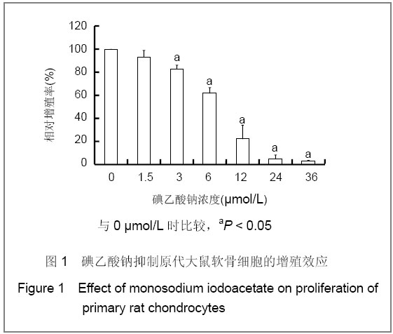
图1 碘乙酸钠抑制原代大鼠软骨细胞的增殖效应
Figure 1 Effect of monosodium iodoacetate on proliferation of primary rat chondrocytes
2.2 碘乙酸钠诱导软骨细胞凋亡 不同剂量碘乙酸钠处理软骨细胞24 h,Hoechst 33342/PI染色,荧光显微镜结果显示,凋亡细胞数量呈逐渐增加的趋势,见图2。
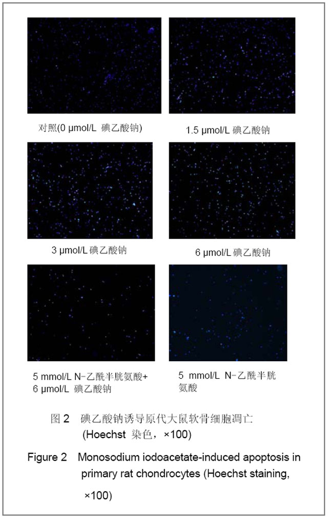
图2 碘乙酸钠诱导原代大鼠软骨细胞凋亡(Hoechst 染色,×100)
Figure 2 Monosodium iodoacetate-induced apoptosis in primary rat chondrocytes (Hoechst staining,×100)
采用FITC-annexin V/PI染色,流式细胞仪分析结果显示,凋亡细胞的增加呈剂量依赖关系,见图3。
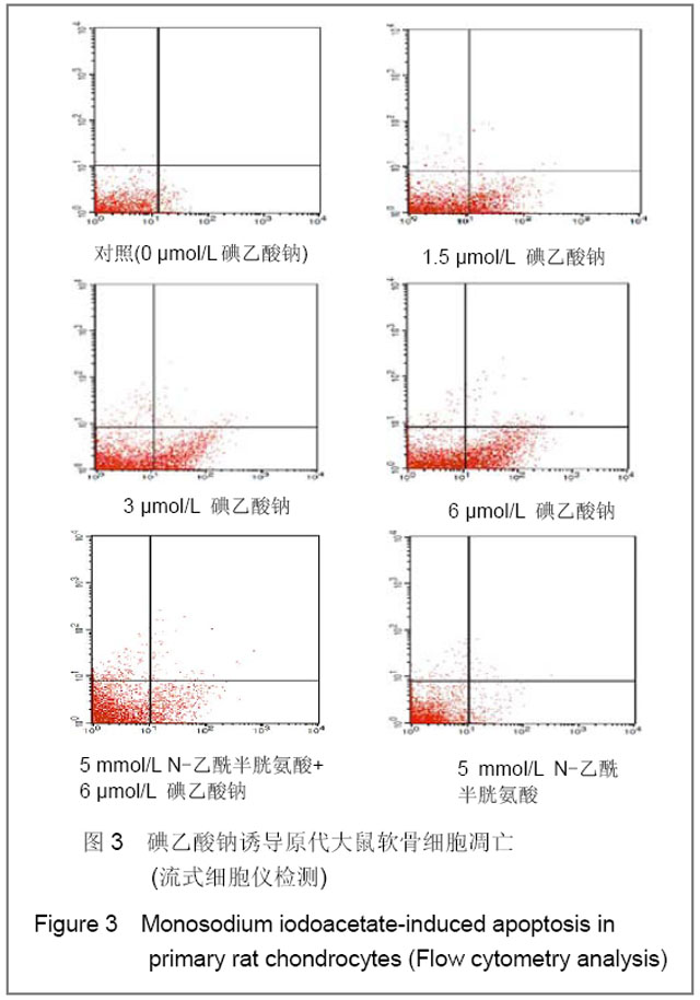
图3 碘乙酸钠诱导原代大鼠软骨细胞凋亡(流式细胞仪检测)
Figure 3 Monosodium iodoacetate-induced apoptosis in primary rat chondrocytes (Flow cytometry analysis)
1.5,3.0,6.0 μmol/L碘乙酸钠处理软骨细胞的凋亡率分别为(17.09±4.18)%,(19.83±1.76)%,(28.00 ±4.25)%,与0 μmol/L碘乙酸钠组(1.31±0.22)%比较差异有显著性意义(P < 0.05)。5 mmol/L N-乙酰半胱氨酸预处理+6.0 μmol/L碘乙酸钠组凋亡率为(16.24±3.60)%,与5 mmol/L N-乙酰半胱氨酸预处理比较差异有显著性意义(P < 0.05),见图4。以上结果说明碘乙酸钠能促进软骨细胞凋亡,呈浓度依赖性,N-乙酰半胱氨酸则能抑制碘乙酸钠诱导的软骨细胞凋亡。
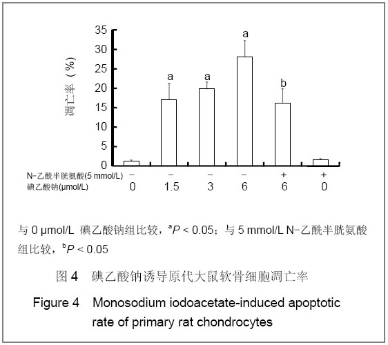
图4 碘乙酸钠诱导原代大鼠软骨细胞凋亡率
Figure 4 Monosodium iodoacetate-induced apoptotic rate of primary rat chondrocytes
2.3 碘乙酸钠诱导软骨细胞活性氧产生 为了探讨碘乙酸钠诱导软骨细胞凋亡形成的机制,采用荧光分光光度计检测碘乙酸钠对细胞内活性氧生成的影响,见图5。细胞荧光强度随碘乙酸钠浓度增大而增高,当碘乙酸钠浓度为6.0 μmol/L时,细胞荧光密度明显增强(
P < 0.05),提示细胞内活性氧水平显著增加。5 mmol/L N-乙酰半胱氨酸预处理+6.0 μmol/L碘乙酸钠组与
6.0 μmol/L碘乙酸钠组比较,活性氧的生成明显减少,其差异有显著性意义(
P < 0.05)。
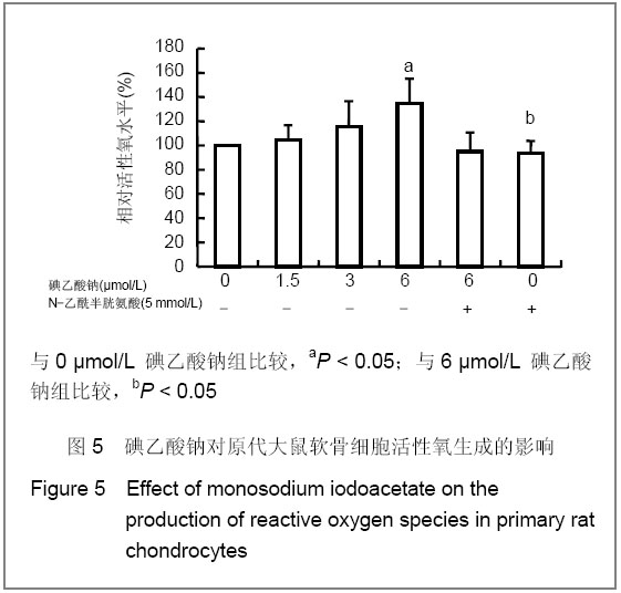
图5 碘乙酸钠对原代大鼠软骨细胞活性氧生成的影响
Figure 5 Effect of monosodium iodoacetate on the production of reactive oxygen species in primary rat chondrocytes
2.4 碘乙酸钠降低软骨细胞线粒体跨膜电位 不同剂量碘乙酸钠处理后的软骨细胞,采用罗丹明123荧光染料染色,荧光分光光度计检测结果显示,线粒体跨膜电位水平随碘乙酸钠浓度增大而下降;当碘乙酸钠浓度为6.0 μmol/L,线粒体跨膜电位明显下降(
P < 0.05)。
5 mmol/L N-乙酰半胱氨酸预处理+6.0 μmol/L碘乙酸钠组与6 μmol/L碘乙酸钠组比较,线粒体跨膜电位的升高具有显著性意义(
P < 0.05),见图6。
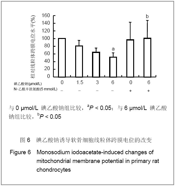
图6 碘乙酸钠诱导软骨细胞线粒体跨膜电位的改变
Figure 6 Monosodium iodoacetate-induced changes of mitochondrial membrane potential in primary rat chondrocytes
同时采用激光扫描共聚焦显微镜观察Rho123沿线粒体膜的分布。随着碘乙酸钠浓度的增加,Rho123在线粒体膜的累积减少,细胞的荧光染色强度逐渐降低,5 mmol/L N-乙酰半胱氨酸预处理+6.0 μmol/L碘乙酸钠组细胞的荧光染色强度明显大于6.0 μmol/L碘乙酸钠组,见图7。结果提示碘乙酸钠致软骨细胞线粒体跨膜电位降低呈剂量依赖性关系,N-乙酰半胱氨酸则能抑制线粒体跨膜电位降低。
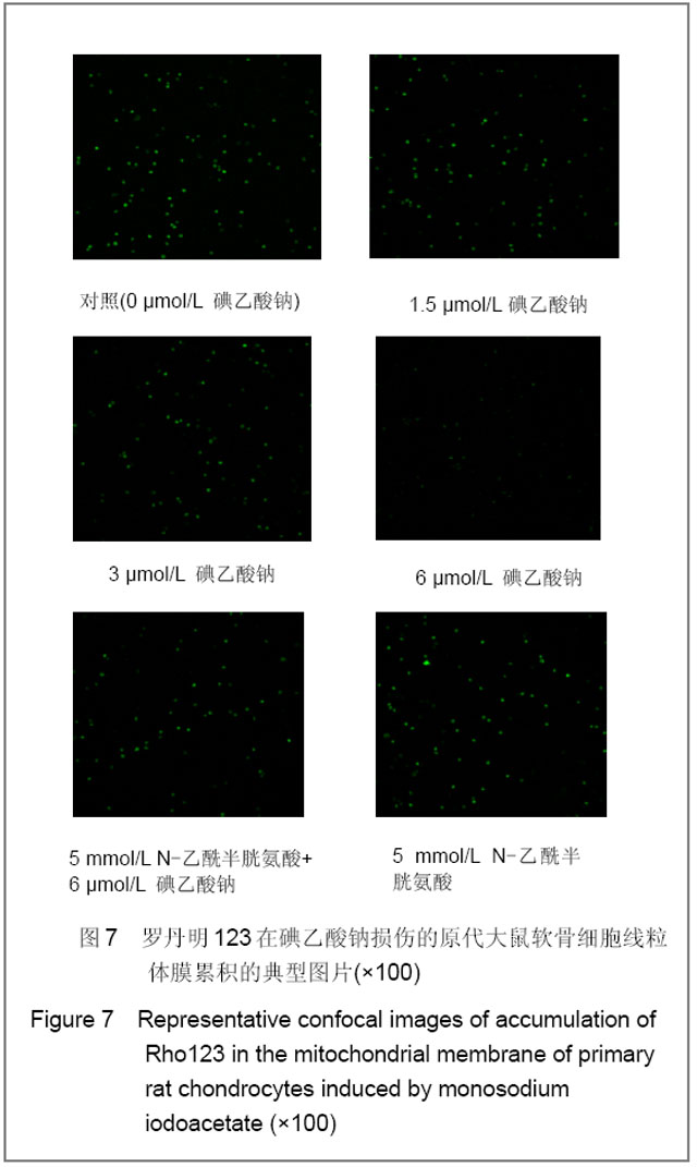
图7 罗丹明123在碘乙酸钠损伤的原代大鼠软骨细胞线粒体膜累积的典型图片(×100)
Figure 7 Representative confocal images of accumulation of Rho123 in the mitochondrial membrane of primary rat chondrocytes induced by monosodium iodoacetate (×100)
2.5 碘乙酸钠诱导细胞色素C的释放和Caspase-3的蛋白表达 为了解碘乙酸钠是否通过细胞色素C的释放启动凋亡,检测不同剂量碘乙酸钠对细胞色素C释放的影响。当碘乙酸钠浓度为3.0和6.0 μmol/L时,细胞色素C蛋白表达与0 μmol/L碘乙酸钠组比较差异均有显著性意义(P < 0.05),见图8A。此外,碘乙酸钠处理后的细胞,caspase-3蛋白表达呈剂量依赖式增加,与0 μmol/L碘乙酸钠组比差异均有显著性意义(P < 0.05),见图8B。N-乙酰半胱氨酸能够有效抑制细胞色素C的释放(P < 0.05)和caspase-3的蛋白表达(P < 0.05)。
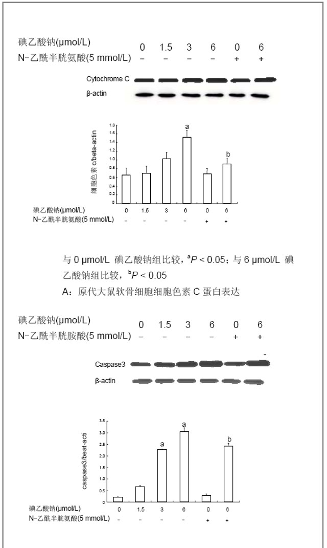
图8 碘乙酸钠对原代大鼠软骨细胞caspase-3和细胞色素C蛋白表达水平的影响
Figure 8 Western blot showing changes in the levels of cytochrome C and caspase-3 in primary rat chondrocytes induced by monosodium iodoacetate


