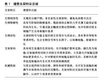
| [1] Ashman O,Phillips AM.Treatment of non-unions with bone defects: which option and why? Injury. 2013;44 Suppl 1:S43-45.[2] Thaller PH,Furmetz J,Wolf F,et al.Limb lengthening with fully implantable magnetically actuated mechanical nails (PHENIX((R)))-preliminary results.Injury.2014;45 Suppl 1:S60-65.[3] Hollister SJ.Porous scaffold design for tissue engineering.Nat Mater.2005;4(7):518-524.[4] Garcia-Gareta E,Coathup MJ,Blunn GW.Osteoinduction of bone grafting materials for bone repair and regeneration.Bone. 2015; 81:112-121.[5] Brydone AS,Meek D,Maclaine S.Bone grafting, orthopaedic biomaterials, and the clinical need for bone engineering.Proc Inst Mech Eng H.2010;224(12):1329-1343.[6] Higgins A,Glover M,Yang Y,et al.EXOGEN ultrasound bone healing system for long bone fractures with non-union or delayed healing: a NICE medical technology guidance. Appl Health Econ Health Policy. 2014;12(5):477-484.[7] Faour O,Dimitriou R,Cousins CA, Giannoudis PV. The use of bone graft substitutes in large cancellous voids: any specific needs? Injury.2011;42 Suppl 2:S87-90.[8] O'Keefe RJ, Mao J. Bone tissue engineering and regeneration: from discovery to the clinic--an overview. Tissue Eng Part B Rev. 2011;17(6):389-392.[9] Bose S,Roy M,Bandyopadhyay A.Recent advances in bone tissue engineering scaffolds.Trends Biotechnol.2012;30(10):546-554.[10] Wu S, Liu X,Yeung KWK,et al. Biomimetic porous scaffolds for bone tissue engineering.Mater Sci Eng R. 2014;80:1-36.[11] Webber MJ,Khan OF,Sydlik SA, et al. R. A perspective on the clinical translation of scaffolds for tissue engineering.Ann Biomed Eng.2015;43(3):641-656.[12] Boskey AL,Roy R.Cell culture systems for studies of bone and tooth mineralization. Chem Rev. 2008;108(11):4716-4733.[13] Young MF.Bone matrix proteins: their function, regulation, and relationship to osteoporosis. Osteoporos Int.2003;14 Suppl 3: S35-42.[14] Kim SS,Sun Park M,Jeon O,et al. Poly(lactide-co-glycolide)/ hydroxyapatite composite scaffolds for bone tissue engineering. Biomaterials.2006;27(8):1399-1409.[15] Hench LL.Opening paper 2015- Some comments on Bioglass: Four Eras of Discovery and Development. Biomed Glasses. 2015;1(1):1-11.[16] Albrektsson T,Johansson C. Osteoinduction, osteoconduction and osseointegration. Eur Spine J. 2001;10 Suppl 2:S96-101.[17] Vaccaro AR.The role of the osteoconductive scaffold in synthetic bone graft. Orthopedics. 2002;25(5 Suppl):s571-578.[18] Gao C, Deng Y,Feng P, et al.Current progress in bioactive ceramic scaffolds for bone repair and regeneration. Int J Mol Sci. 2014; 15(3):4714-4732.[19] Loh QL, Choong C. Three-dimensional scaffolds for tissue engineering applications: role of porosity and pore size.Tissue Eng Part B Rev.2013;19(6):485-502.[20] Johnson T, Bahrampourian R,Patel A,et al.Fabrication of highly porous tissue-engineering scaffolds using selective spherical porogens.Biomed Mater Eng.2010;20(2):107-118.[21] Dehghani F, Annabi N. Engineering porous scaffolds using gas-based techniques. Curr Opin Biotechnol. 2011;22(5):661-666.[22] Huang Y, Han S, Pang X,et al. Electrodeposition of porous hydroxyapatite/calcium silicate composite coating on titanium for biomedical applications.Appl Surf Sci.2013;271:299-302.[23] Ligon SC,Liska R,Stampfl J,et al. Polymers for 3D Printing and Customized Additive Manufacturing. Chem Rev. 2017;117(15): 10212-10290.[24] Kim K,Yeatts A,Dean D,et al.Stereolithographic bone scaffold design parameters: osteogenic differentiation and signal expression.Tissue Eng Part B Rev.2010;16(5):523-539.[25] Do AV, Khorsand B, Geary SM, et al. 3D Printing of Scaffolds for Tissue Regeneration Applications. Adv Healthc Mater. 2015;4(12): 1742-1762.[26] Xia Y,Zhou P,Cheng X,et al.Selective laser sintering fabrication of nano-hydroxyapatite/poly-epsilon-caprolactone scaffolds for bone tissue engineering applications. Int J Nanomedicine. 2013;8: 4197-4213.[27] Murphy SV,Atala A.3D bioprinting of tissues and organs.Nat Biotechnol.2014;32(8):773-785.[28] Saunders RE, Derby B. Inkjet printing biomaterials for tissue engineering: bioprinting. Int Mater Rev.2014;59(8):430-448.[29] Koch L,Gruene M,Unger C,et al.Laser assisted cell printing.Curr Pharm Biotechnol. 2013;14(1):91-97.[30] Faulkner-Jones A,Fyfe C,Cornelissen DJ,et al.Bioprinting of human pluripotent stem cells and their directed differentiation into hepatocyte-like cells for the generation of mini-livers in 3D. Biofabrication.2015;7(4):044102.[31] Ozbolat IT, Hospodiuk M. Current advances and future perspectives in extrusion-based bioprinting. Biomaterials. 2016;76:321-343.[32] Staiger MP,Pietak AM,Huadmai J, et al.Magnesium and its alloys as orthopedic biomaterials: a review. Biomaterials. 2006; 27(9): 1728-1734.[33] Lopez-Heredia MA,Sohier J,Gaillard C,et al.Rapid prototyped porous titanium coated with calcium phosphate as a scaffold for bone tissue engineering. Biomaterials.2008;29(17):2608-2615.[34] Chen H,Wang C,Zhu X,et al.Fabrication of porous titanium scaffolds by stack sintering of microporous titanium spheres produced with centrifugal granulation technology. Mater Sci Eng C Mater Biol Appl.2014;43:182-188.[35] Bose S,Fielding G,Tarafder S,et al.Understanding of dopant-induced osteogenesis and angiogenesis in calcium phosphate ceramics.Trends Biotechnol.2013;31(10):594-605.[36] Atkins GJ,Welldon KJ,Halbout P,et al.Strontium ranelate treatment of human primary osteoblasts promotes an osteocyte-like phenotype while eliciting an osteoprotegerin response. Osteoporos Int.2009;20(4):653-664.[37] Lei Y,Xu Z,Ke Q,et al.Strontium hydroxyapatite/chitosan nanohybrid scaffolds with enhanced osteoinductivity for bone tissue engineering. Mater Sci Eng C Mater Biol Appl. 2017;72: 134-142.[38] Xiu P,Jia ZJ,Lv J, et al.Hierarchical Micropore/Nanorod Apatite Hybrids In-Situ Grown from 3-D Printed Macroporous Ti6Al4V Implants with Improved Bioactivity and Osseointegration.J Mater Sci Technol.2017;33(2):179-186.[39] Peuster M,Hesse C,Schloo T,et al.Long-term biocompatibility of a corrodible peripheral iron stent in the porcine descending aorta. Biomaterials.2006;27(28):4955-4962.[40] Chou DT,Wells D,Hong D,et al. Novel processing of iron-manganese alloy-based biomaterials by inkjet 3-D printing. Acta Biomater.2013;9(10):8593-8603.[41] Ho GW, Matinlinna JP.Insights on Ceramics as Dental Materials. Part I: Ceramic Material Types in Dentistry. Silicon. 2011;3(3): 109-115.[42] Bohner M, Galea L, Doebelin N. Calcium phosphate bone graft substitutes: Failures and hopes. J Eur Ceram Soc. 2012;32(11): 2663-2671.[43] Ko CL, Chen WC, Chen JC, et al. Properties of osteoconductive biomaterials: calcium phosphate cement with different ratios of platelet-rich plasma as identifiers. Mater Sci Eng C Mater Biol Appl.2013;33(6):3537-3544.[44] Tang Z,Tan Y,Ni Y,et al.Comparison of ectopic bone formation process induced by four calcium phosphate ceramics in mice. Mater Sci Eng C Mater Biol Appl.2017;70(Pt 2):1000-1010.[45] Mohseni M,Jahandideh A,Abedi G,et al.Assessment of tricalcium phosphate/collagen (TCP/collagene)nanocomposite scaffold compared with hydroxyapatite (HA) on healing of segmental femur bone defect in rabbits.Artif Cells Nanomed Biotechnol. 2018;46(2):242-249.[46] Tarafder S,Bose S.Polycaprolactone-coated 3D printed tricalcium phosphate scaffolds for bone tissue engineering: in vitro alendronate release behavior and local delivery effect on in vivo osteogenesis.ACS Appl Mater Interfaces.2014;6(13):9955-9965.[47] Dai Y,Liu H,Liu B,et al.Porous β-Ca2SiO4 ceramic scaffolds for bone tissue engineering: In vitro and in vivo characterization. Ceram Int.2015;41(4):5894-5902.[48] Pasold J,Markhoff J,Tillmann J,et al.Direct influence of titanium and zirconia particles on the morphology and functionality of mature human osteoclasts. J Biomed Mater Res A. 2017; 105(9): 2608-2615.[49] Jones JR.Review of bioactive glass: from Hench to hybrids. Acta Biomater.2013;9(1):4457-4486.[50] Rahaman MN,Day DE,Bal BS,et al.Bioactive glass in tissue engineering.Acta Biomater.2011;7(6):2355-2373.[51] Miguez-Pacheco V,Hench LL,Boccaccini AR.Bioactive glasses beyond bone and teeth: emerging applications in contact with soft tissues. Acta Biomater.2015;13:1-15.[52] Habraken WJ, Wolke JG, Jansen JA. Ceramic composites as matrices and scaffolds for drug delivery in tissue engineering.Adv Drug Deliv Rev.2007;59(4-5):234-248.[53] Westhauser F,Weis C,Prokscha M,et al. Three-dimensional polymer coated 45S5-type bioactive glass scaffolds seeded with human mesenchymal stem cells show bone formation in vivo. J Mater Sci Mater Med. 2016;27(7):119.[54] Jiang S, Zhang Y, Shu Y, et al.Amino-functionalized mesoporous bioactive glass for drug delivery. Biomed Mater. 2017; 12(2): 025017.[55] Zhang Y,Cui X,Zhao S, et al. Evaluation of injectable strontium-containing borate bioactive glass cement with enhanced osteogenic capacity in a critical-sized rabbit femoral condyle defect model.ACS Appl Mater Interfaces. 2015;7(4): 2393-2403.[56] Ravi S, Chaikof EL. Biomaterials for vascular tissue engineering. Regen Med. 2010;5(1):107-120.[57] Zhu J,Marchant RE. Design properties of hydrogel tissue-engineering scaffolds. Expert Rev Med Devices. 2011;8(5): 607-626.[58] Sheikh FA, Ju HW, Moon BM, et al. Hybrid scaffolds based on PLGA and silk for bone tissue engineering. J Tissue Eng Regen Med.2016;10(3):209-221.[59] Liao HT, Chen CT, Chen JP. Osteogenic differentiation and ectopic bone formation of canine bone marrow-derived mesenchymal stem cells in injectable thermo-responsive polymer hydrogel. Tissue Eng Part C Methods.2011;17(11):1139-1149.[60] Woodruff MA, Hutmacher DW. The return of a forgotten polymer - Polycaprolactone in the 21st century. Prog Polym Sci. 2010; 35(10):1217-1256.[61] Hollister SJ, Murphy WL. Scaffold translation: barriers between concept and clinic.Tissue Eng Part B Rev.2011;17(6):459-474.[62] Gloria A,De Santis R,Ambrosio L. Polymer-based composite scaffolds for tissue engineering. J Appl Biomater Biomech. 2010; 8(2):57-67.[63] Bellis SL. Advantages of RGD peptides for directing cell association with biomaterials. Biomaterials. 2011;32(18): 4205-4210.[64] Villa MM,Wang L,Huang J,et al.Bone tissue engineering with a collagen-hydroxyapatite scaffold and culture expanded bone marrow stromal cells. J Biomed Mater Res B Appl Biomater. 2015; 103(2):243-253.[65] Marcacci M,Kon E,Moukhachev V,et al.Stem cells associated with macroporous bioceramics for long bone repair: 6- to 7-year outcome of a pilot clinical study.Tissue Eng.2007;13(5):947-955.[66] Hou J,Wang J,Cao LY,et al.Segmental bone regeneration using rhBMP-2-loaded collagen/chitosan microspheres composite scaffold in a rabbit model.Biomed Mater.2012;7(3):035002.[67] Chang YS,Huang HD,Yeh KT, et al.Genetic alterations in endometrial cancer by targeted next-generation sequencing.Exp Mol Pathol.2016;100(1):8-12.[68] Yang T,Hu Y,Wang CY,et al.Fabrication of Hierarchical Macroporous Biocompatible Scaffolds by Combining Pickering High Internal Phase Emulsion Templates with Three-Dimensional Printing. ACS Appl Mater Interfaces.2017;9(27):22950-22958.[69] Shuai CJ,Yang B,Peng SP,et al.Development of composite porous scaffolds based on poly(lactide-co-glycolide)/ nano-hydroxyapatite via selective laser sintering. Int J Adv Manuf Tech. 2013;69(1-4):51-7.[70] Haider A, Gupta KC, Kang IK. Morphological effects of HA on the cell compatibility of electrospun HA/PLGA composite nanofiber scaffolds. Biomed Res Int.2014;2014:308306. |
.jpg)

.jpg)