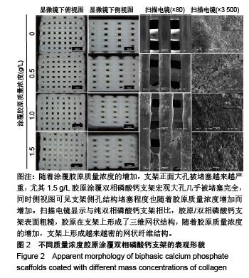
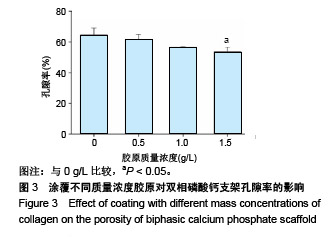
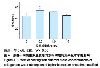
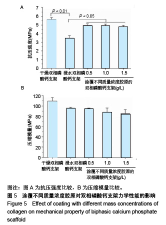
| [1]Nerem RM,Sambanis A.Tissue engineering: from biology to biological substitutes. Tissue Eng.1995;1(1):3-13.[2]Li S,Li N.Effects of composition and temperature on porosity and pore size distribution of porous ceramics prepared from Al (OH) 3, and kaolinite gangue.Ceram Int. 2007;33(4):551-556.[3]Chen J,Wang Y,Chen X,et al.Biomimetic synthesis of porous composite scaffold composed of nano-hydroxyapatite and chitosan by freeze-drying technology.Rare Metal Mat Eng. 2009;38(12): 271-273.[4]Zhao K,Wei J,Luo D,et al.Fabrication of hydroxyapatite porous scaffolds by freeze drying.J Chin Ceram Soc.2009;37(3):432-435. [5]Sepulveda P,Binner JG,Rogero SO,et al.Production of porous hydroxyapatite by the gel-casting of foams and cytotoxic evaluation. J Biomed Mater Res.2000;50(1):27-34.[6]Chia HN,Wu BM.Recent advances in 3D printing of biomaterials. J Biol Eng.2015;9(1):4. [7]Webb PA.A review of rapid prototyping (RP) techniques in the medical and biomedical sector.J Med Eng Technol.2000;24(4):149-153.[8]Duoss EB,Twardowski M,Lewis JA.Sol-Gel inks for direct-write assembly of functional oxides.Adv Mater.2007;19(21):3485-3489.[9]尤琦,张赢心,李佳乐,等.双相磷酸钙陶瓷化学组成对其材料性能的影响[J].海南医学,2016, 27(15):2502-2504. [10]许瑾,吴晶晶,王晓冬,等.羟基磷灰石复合骨组织工程支架的研究进展[J].生物骨科材料与临床研究,2016,13(2):63-66. [11]Faruq O,Kim B,Padalhin AR,et al.A hybrid composite system of biphasic calcium phosphate granules loaded with hyaluronic acid-gelatin hydrogel for bone regeneration. J Biomater Appl. 2017;32(4):433-445.[12]Castilho M,Moseke C,Ewald A,et al.Direct 3D powder printing of biphasic calcium phosphate scaffolds for substitution of complex bone defects.Biofabrication.2014;6(1): 015006.[13]Ng A MH,Tan KK,Phang MY,et al.Differential osteogenic activity of osteoprogenitor cells on HA and TCP/HA scaffold of tissue engineered bone.J Biomed Mater Res.2008;85A(2):301-312. [14]Ebrahimi M,Botelho M.Biphasic calcium phosphates (BCP) of hydroxyapatite (HA) and tricalcium phosphate (TCP) as bone substitutes: importance of physicochemical characterizations in biomaterials studies.Data Brief.2017;10(C):93-97.[15]Kim JW,Shin KH,Koh YH,et al.Production of poly(ε-caprolactone)/ hydroxyapatite composite scaffolds with a tailored macro/micro- porous structure, high mechanical properties, and excellent bioactivity.Materials.2017;10(10):1123-1135. [16]王庆利,何丹,郑晓冬,等.有机溶剂对细胞的毒害及细胞的耐受性机制[J].微生物学通报, 2002,29(4):81-85. [17]Stratton S,Shelke NB,Hoshino K,et al.Bioactive polymeric scaffolds for tissue engineering.Bioact Mater.2016;1(2):93-108.[18]李涤尘,连芩,靳忠民,等.基于光固化和凝胶注模的镁合金/生物陶瓷骨支架及其成型方法:CN 102335460 A[P].2012.[19]Zhang D,Wu X,Chen J,et al.The development of collagen based composite scaffolds for bone regeneration.Bioact Mater. 2018; 3(1):129-138.[20]Lim MM,Sun T,Sultana N.In vitro biological evaluation of electrospun polycaprolactone/gelatine nanofibrous scaffold for tissue engineering. J Nanomater.2015;(2):1-10.[21]Bostman OM,Pihlajamaki HK.Adverse tissue reactions to bioabsorbable: Saos-2 are influenced by collagen fibers in xenogenic bone biomaterial: fixation devices.Clin Orthop Relat Res. 2000;(371):216-227.[22]臧晓龙,孙健,李亚莉,等.3D生物打印构建聚乳酸羟基乙酸/纳米羟基磷灰石支架骨形态发生蛋白2缓释复合体的实验研究[J].中国组织工程研究,2016,20(16):2405-2411.[23]马艺娟.三维可控微结构多孔生物活性陶瓷的制备及其性能研究[D].华南理工大学, 2015. [24]Lee MH,You C,Kim KH.Combined effect of a microporous layer and type I collagen coating on a biphasic calcium phosphate scaffold for bone tissue engineering.Materials.2015; 8(3):1150-1161. [25]Takeuchi Y,Nakayama K,Matsumoto T.Differentiation and cell surface expression of transforming growth factor-beta receptors are regulated by interaction with matrix collagen in murine osteoblastic cells.J Biol Chem.1996;271(7):3938-3944.[26]Mizuno M,Fujisawa R,Kuboki Y.Type I collagen‐induced osteoblastic differentiation of bone‐marrow cells mediated by collagen‐α2β1 integrin interaction.J Cell Physiol. 2000;184(2): 207-213. [27]Roehlecke C, Witt M, Kasper M,et al.Synergistic effect of titanium alloy and collagen type I on cell adhesion, proliferation and differentiation of osteoblast-like cells. Cells Tissues Organs. 2001; 168(3):178-187.[28]Dorozhkin SV. Calcium orthophosphate-based biocomposites and hybrid biomaterials. J Mater Sci.2009;44(9):2343-2387.[29]Wei QR,Lu J,Zhao HC,et al.Preparation and characterization of the microspheres comprised of atelocollagen and BCP.Key Eng Mater. 2007;330-332:423-426.[30]Ebrahimi M,Pripatnanont P,Monmaturapoj N,et al.Fabrication and characterization of novel nano hydroxyapatite/β-tricalcium phosphate scaffolds in three different composition ratios.J Biomed Mater Res.2012;100(9):2260-2268.[31]胡涛.下颌骨三维重建及计算机辅助低温沉积制造研究[D].杭州:杭州电子科技大学,2015.[32]Suo H,Xu K,Zheng X.Using glucosamine to improve the properties of photocrosslinked gelatin scaffolds.J Biomater Appl. 2015;29(7): 977-987. [33]Zheng H, Bai Y, Shih MS,et al.Effect of a β-TCP collagen composite bone substitute on healing of drilled bone voids in the distal femoral condyle of rabbits.J Biomed Mater Res.2014;102(2):376-383.[34]Curran JM,Gallagher JA,Hunt JA.The inflammatory potential of biphasic calcium phosphate granules in osteoblast/macrophage co-culture.Biomaterials.2005;26(26): 5313-5320. [35]Lin F,Liao C,Chen K,et al.Preparation of β-TCP/HAP biphasic ceramics with natural bone structure by heating bovine cancellous bone with the addition of (NH4)2HPO4.J Biomed Mater Res. 2000; 51(2):157-163.[36]孙开瑜,徐铭恩,周永勇.基于3D 打印的 I 型胶原涂覆 β-TCP 骨组织工程支架研究[J]. 中国生物医学工程学报,2018,37(3):335-343.[37]Morra M Cassinelli C,Cascardo G,et al.Surface engineering of titanium by collagen immobilization. Surface characterization and in vitro and in vivo studies. Biomaterials.2003;24(25):4639-4654.[38]Henkel J,Woodruff MA,Epari DR,et al.Bone regeneration based on tissue engineering conceptions-a 21st century perspective.Bone Res.2013;1(3):216-248.[39]Olszta MJ,Cheng X,Sang SJ,et al.Bone structure and formation: a new perspective. Mater Sci Eng R.2007;58(3):77-116.[40]Wang Y,Wang K,Li X,et al.3D fabrication and characterization of phosphoric acid scaffold with a HA/β-TCP weight ratio of 60:40 for bone tissue engineering applications.PLoS One.2017;12(4):e0174870.[41]尤琦.三维打印技术构建双相磷酸钙陶瓷支架及其性能研究[D].长春:吉林大学,2016.[42]袁景,甄平,赵红斌.高性能多孔β-磷酸三钙骨组织工程支架的3D打印[J].中国组织工程研究,2014,18(43):6914-6921. [43]Andrew G,Dafydd V,Jing Y,et al.Review of additive manufactured tissue engineering scaffolds: relationship between geometry and performance.Burns Trauma.2018;6(1):19.[44]Pilliar RM,Filiaggi MJ,Wells JD,et al.Porous calcium polyphosphate scaffolds for bone substitute applications-in vitro characterization. Biomaterials.2001;22(9): 963-972.[45]Ma Z,Mao Z,Gao C.Surface modification and property analysis of biomedical polymers used for tissue engineering.Colloids Surf B Biointerfaces.2007;60(2):137-157. [46]Daculsi G,Legeros R.Biphasic Calcium Phosphate (BCP) bioceramics: chemical, physical, and biological properties.Encycl Biomater Biomed Eng.2008:359-366. [47]LeGeros RZ,Lin S,Rohanizadeh R,et al.Biphasic calcium phosphate bioceramics: preparation, properties and applications.J Mater Sci Mater Med.2003;14(3):201-209. |
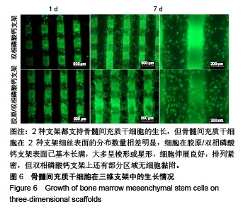
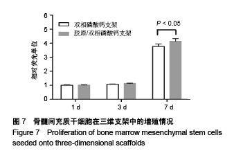
.jpg)


.jpg)