中国组织工程研究 ›› 2019, Vol. 23 ›› Issue (4): 562-566.doi: 10.3969/j.issn.2095-4344.1037

• 数字化骨科 digital orthopedics • 上一篇    下一篇

困难气道患者三维有限元模型解剖差异的分析

谢雨晨,陈文栋,马 莉   

  1. 昆明医科大学第一附属医院麻醉科,云南省昆明市 650032
  • 出版日期:2019-02-08 发布日期:2019-02-08
  • 通讯作者: 陈文栋,主治医师,昆明医科大学第一附属医院麻醉科,云南省昆明市 650032
  • 作者简介:谢雨晨,女,1991年生,汉族,山东省菏泽市人,昆明医科大学在读硕士,主要从事围术期困难气道的评估研究。
  • 基金资助:

    云南省科技厅内设研究机构资助项目(2014NS083),项目负责人:陈文栋;云南省应用基础研究(昆医联合专项)[2017FE468(-147) ],项目负责人:陈文栋

Anatomical differences in three-dimensional finite element model of difficult airway patients

Xie Yuchen, Chen Wendong, Ma Li   

  1. Department of Anesthesiology, First Affiliated Hospital of Kunming Medical University, Kunming 650032, Yunnan Province, China
  • Online:2019-02-08 Published:2019-02-08
  • Contact: Chen Wendong, Attending physician, Department of Anesthesiology, First Affiliated Hospital of Kunming Medical University, Kunming 650032, Yunnan Province, China
  • About author:Xie Yuchen, Master candidate, Department of Anesthesiology, First Affiliated Hospital of Kunming Medical University, Kunming 650032, Yunnan Province, China
  • Supported by:

    the Research Project of Yunnan Provincial Science and Technology Department, No. 2014NS083 (to CWD); the Applied Basic Research Project of Yunnan Province (Combined Project of Kunming Medical University), No. 2017FE468(-147) (to CWD)

摘要:

文章快速阅读:


文题释义:
三维实体建模:是一种集计算机辅助绘图与参数化实体造型、装配造型、二维和三维双向关联绘图及图形转换器等模块为一体的现代机械设计手段。实验运用Mimics软件对正常人体上气道创建三维实体模型,并通过mimics软件自带测量工具进行数据测量,分析该模型在临床工作中的实用性,可为临床工作提供数据分析,指导临床工作。
困难气道:是指可能对麻醉后维护气道通畅及气管插管造成困难的各种临床情况,包括面罩给氧困难和气管内插管困难。研究通过对正常气道及困难气道三维模型进行数据测量,对其在平卧位及嗅花位变化时口咽及上气道的数值变化进行解剖分析,对临床工作具有重要意义。
 
摘要
背景:近年来随着科学技术的发展,三维模型越来越多的运用到医学领域中。气道管理是麻醉手术中最重要环节之一,而在国内外较少见有关人体气道三维有限元模型的研究。
目的:应用MRI检查获得的二维断层图像重建人体气道三维有限元模型,分析困难气道三维模型的解剖差异。
方法:纳入手术麻醉过程中确定为正常气道和困难气道患者各20例,获取其头颈部核磁共振二维图像数据,应用Mimics10.01软件重建包括整个下颌骨、舌骨及上气道等在内的三维实体数字化模型,利用Mimics自带测量软件测量相关指标,同时对正常人体体表标志进行测量并获得相关数据,与模型所测指标行配对比较,分析困难气道患者与正常气道患者的解剖差异。
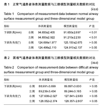
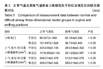
结果与结论:①正常气道患者下颌距离的三维模型组测量值较正常人体体表测量组偏小(P < 0.01);困难气道患者下颌距离的三维模型组测量值较正常人体体表测量组偏大,但差异无统计学意义(P > 0.05);正常气道、困难气道患者下颌角角度的三维模型组测量值较正常人体体表测量组偏大,但差异无统计学意义(P > 0.05);②在平卧位及嗅花位时,正常气道组口咽角角度、矢状切最小横截面积均大于与困难气道组(P < 0.05),两组冠状切最小横截面积比较差异无显著性意义(P > 0.05);③结果表明,运用 MRI人体气道数据可建立较为真实可靠的三维有限元模型,利用三维有限元模型可有效分析困难气道患者的解剖差异,为以后临床上工作中预测困难气道提供参考。

中国组织工程研究杂志出版内容重点:人工关节;骨植入物;脊柱骨折;内固定;数字化骨科;组织工程
ORCID: 0000-0002-4521-4825(谢雨晨)

关键词: 困难气道, 气道管理, MRI, 三维有限元模型, 气道解剖差异

Abstract:

BACKGROUND: Three-dimensional models are more and more applied in medical fields with technology development. Airway management is a key for anesthesia; however, litter is reported on the three-dimensional finite element models of normal human airways.

OBJECTIVE: To reconstruct the three-dimensional finite element model of human airway based on MRI data, and to analyze the anatomical differences in difficult airway three-dimensional model.
METHODS: Twenty patients with normal airways and 20 patients with difficult airways were enrolled, and then the MRI two-dimensional imaging data of head and neck were obtained. A three-dimensional solid digital model including the entire mandible, hyoid bone and upper airway was reconstructed using Mimics 10.01. Measurement software was used to measure related indicators. At the same time, normal human body surface markers were measured to obtain relevant data to compare with the measured indexes of the model, and the differences in normal and difficult airway anatomy were analyzed.
RESULTS AND CONCLUION: (1) The measured value of mandibular distance in the three-dimensional model group was significantly smaller than that in the body surface measurement group in normal airway patients (P < 0.01). The measured value of mandibular distance in the three-dimensional model group was higher than that in the body surface measurement group in patients with difficult airway (P > 0.05). The measured value of mandibular angle in the three-dimensional model group was higher than that in the body surface measurement group of normal airway patients (P > 0.05). (2) In supine and sniffing positions, the oropharyngeal angle and minimal sagittal cross-sectional area in the normal airway group were significantly higher than those in the difficult airway group (P < 0.05). There was no significant difference in the minimal coronal cross-sectional area between two groups (P > 0.05). (3) These results suggest that the human airway data obtained by MRI can establish a realistic and reliable three-dimensional finite element model, and the model can accurately analyze the anatomical differences with difficult airways, which provides reference for predicting difficult airways in clinical practice. 

中国组织工程研究杂志出版内容重点:人工关节;骨植入物;脊柱骨折;内固定;数字化骨科;组织工程

Key words: Anesthesia, Airway Management, Magnetic Resonance Imaging, Finite Element Analysis, Tissue Engineering

中图分类号: