中国组织工程研究 ›› 2019, Vol. 23 ›› Issue (3): 421-426.doi: 10.3969/j.issn.2095-4344.1025

• 组织构建实验造模 experimental modeling in tissue construction • 上一篇    下一篇

骨性关节炎与骨质疏松症共病模型兔的建立及鉴定

马文娟1,张开伟2,沈冯君2,邓致远3,程效进4   

  1.  (1金沙县中医院骨伤科,贵州省金沙县  551800;2贵州省中医院骨伤科,贵州省贵阳市  550000;3武汉市中医院疼痛科,湖北省武汉市  430050;4山东省曹县中医院,山东省曹县  274400)
  • 收稿日期:2018-08-07
  • 通讯作者: 张开伟,博士,主任医师,贵州省中医院骨伤科,贵州省贵阳市 550000
  • 作者简介:马文娟,1987年生,汉族,湖南省人,2017年贵阳中医学院毕业,硕士,主要从事骨与关节退行性改变及运动损伤研究。
  • 基金资助:

    贵州省优秀科技教育人才省长专项资金(黔省专合字[2012]34号),项目负责人:张开伟;贵州省科技厅项目(黔科合中药字[2011]LKZ7022号),项目负责人:张开伟

Establishment and identification of a rabbit model of comorbid osteoarthritis and osteoporosis

Ma Wenjuan1, Zhang Kaiwei2, Shen Fengjun2, Deng Zhiyuan3, Cheng Xiaojin4   

  1.  (1Jinsha Country Hospital of Traditional Chinese Medicine, Jinsha 551800, Guizhou Province, China; 2Guizhou Provincial Hospital of Traditional Chinese Medicine, Guiyang 550000, Guizhou Province, China; 3Department of Pain, Wuhan Hospital of Traditional Chinese Medicine, Wuhan 430050, Hubei Province, China; 4Caoxian Country Hospital of Traditional Chinese Medicine, Caoxian 274400, Shandong Province, China)
  • Received:2018-08-07
  • Contact: Zhang Kaiwei, MD, Chief physician, Guizhou Provincial Hospital of Traditional Chinese Medicine, Guiyang 550000, Guizhou Province, China
  • About author:Ma Wenjuan, Master, Jinsha Country Hospital of Traditional Chinese Medicine, Jinsha 551800, Guizhou Province, China
  • Supported by:

    Governor’s Fund for Excellent Technological and Educational Talents in Guizhou Province, No. [2012]34 (to ZKW); the Project of Guizhou Provincial Science and Technology Department, No. [2011]LKZ7022 (to ZKW)

摘要:

文章快速阅读:

文题释义:
骨性关节炎:增生性骨关节病是指由于关节退行性变,以致关节软骨被破坏而引起的慢性关节病,又称退化性关节炎、骨关节炎及肥大性关节炎等。
骨质疏松症:是多种原因引起的一组骨病,骨组织有正常的钙化,钙盐与基质呈正常比例,以单位体积内骨组织量减少为特点的代谢性骨病变。
摘要
背景:
老年人群多同时患有骨性关节炎和骨质疏松症,但目前骨性关节炎与骨质疏松症两者之间的关系尚未明确。
目的:构建骨性关节炎+骨质疏松症动物模型并进行鉴定。
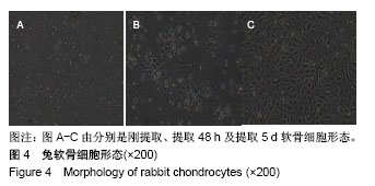
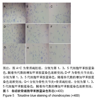
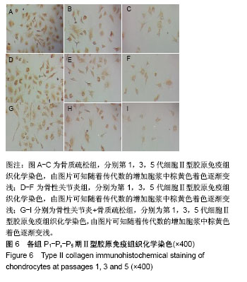
方法:贵阳医学院动物实验中心提供的37只2月龄雌性新西兰大白兔,30只随机分为骨性关节炎组、骨质疏松症组、骨性关节炎+骨质疏松症组,另设7只兔为正常对照组。分别制备骨质疏松症(造模方法:切除双侧卵巢+肌注甲泼尼龙)、骨性关节炎(造模方法:切除前交叉韧带)、骨性关节炎+骨质疏松症(造模方法:切除双侧卵巢+肌注甲泼尼龙+切除前交叉韧带)动物模型,确认造模成功后对上述3种模型软骨进行分离及培养,比较3者软骨细胞中Ⅱ型胶原酶分泌情况。
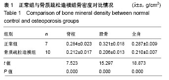
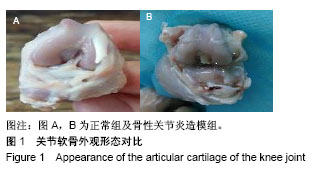
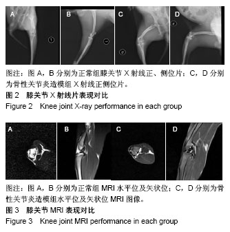
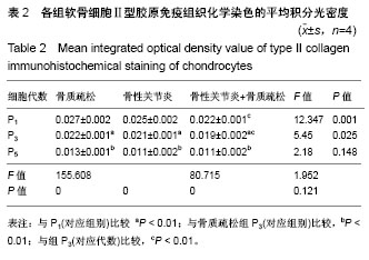
结果与结论:①运用切除兔前交叉韧带、卵巢及肌注甲泼尼松龙琥珀酸钠的方法可使骨质疏松兔骨密度降低25%以上,从关节外观可见明显关节软骨剥脱,膝关节X射线检查可见明显骨赘生成,MRI检测可显示明显关节软骨损伤,由此可知通过上述方法可成功制备骨质疏松症合并骨性关节炎的模型;②骨性关节炎组、骨质疏松症组、骨性关节炎+骨质疏松症3组兔膝关节软骨中均能成功提取出软骨细胞,与骨质疏松症组比较骨性关节炎+骨质疏松组软骨细胞Ⅱ型胶原酶的能力下降差异明显(P < 0.05);③结果说明:实验成功建立了骨性关节炎+骨质疏松症动物模型。

中国组织工程研究杂志出版内容重点:组织构建;骨细胞;软骨细胞;细胞培养;成纤维细胞;血管内皮细胞;骨质疏松组织工程
ORCID: 0000-0001-9397-7408(马文娟)

关键词: 骨性关节炎, 骨质疏松, 动物模型, 前交叉韧带, 卵巢, 关节软骨, 甲泼尼龙, Ⅱ型胶原酶, 免疫组织化学染色, 细胞培养, 组织工程

Abstract:

BACKGROUND: The elderly often suffers from comorbid osteoarthritis and osteoporosis, but the relationship between osteoarthritis and osteoporosis is not yet clear.
OBJECTIVE: To establish and identify an animal model of comorbid osteoarthritis and osteoporosis.
METHODS: Thirty-seven female New Zealand white rabbits at 2 months of age were provided by the Animal Center of Guiyang Medical University, China. Of the 37 rabbits, 30 rabbits were selected and divided into osteoarthritis group, osteoporosis group, osteoarthritis and osteoporosis group. The remaining seven rabbits were used as normal control group. Different animal models of osteoporosis (excision of bilateral ovarian and intramuscular injection of methylprednisolone)/osteoarthritis (excision of anterior cruciate ligament)/osteoarthritis and osteoporosis (excision of bilateral ovaries and anterior cruciate ligament+intramuscular injection of methylprednisolone) were made in corresponding groups. After successful modeling, cartilage samples from each rabbit were isolated and cultured. Secretion of type II collagenase was compared among three groups.
RESULTS AND CONCLUSION: (1) After excision of rabbit anterior cruciate ligament and bilateral ovaries and intramuscular injection of methylprednisolone, the bone density of osteporotic rabbits was reduced by more than 25%. Exfoliation of the articular cartilage could be observed from the joint appearance, and osteophyte formed markedly shown on the X-ray film of the knee joint obviously. Obvious articular cartilage damage was also observed on the MRI image. Therefore, the model of comorbid osteoarthritis and osteoporosis was successfully prepared by the above methods. (2) Chondrocytes could be successfully extracted from the rabbit knee joint cartilage in the three model groups. The ability of chondrocytes secreting type II collagenase was significantly lowered in the osteoporosis group compared to the osteoarthritis and osteoporosis group (P < 0.05). (3) To conclude, the animal model of comorbid osteoarthritis and osteoporosis was successfully established.

中国组织工程研究杂志出版内容重点:组织构建;骨细胞;软骨细胞;细胞培养;成纤维细胞;血管内皮细胞;骨质疏松组织工程

Key words: Osteoarthritis, Osteoporosis, Models, Animal, Tissue Engineering

中图分类号: