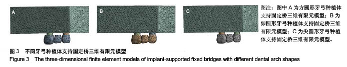
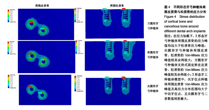
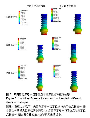
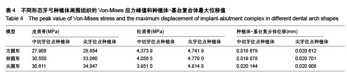
| [1] Romeo E,Lops D,Margutti E,et al.Implant‐supported fixed cantilever prostheses in partially edentulous arches. A seven‐year prospective study.Clin Oral Implants Res. 2003;14(3):303-311.[2] Dede DÖ,Cenk Durmu?lar M,?ah?n O,et al.Telescopic Overdenture and Implant Supported Fixed Partial Denture: A Pragmatic Treatment Approach.Case Rep Dent. 2015;2015:392397.[3] Sagat G,Yalcin S,Gultekin BA,et al.Influence of arch shape and implant position on stress distribution around implants supporting fixed full-arch prosthesis in edentulous maxilla.Implant Dent.2010;19(6):498-508.[4] 袁豪,周延民.上颌All-on-4修复种植体在不同牙弓形态中应力分布的三维有限元分析[J].吉林大学学报,2014,40(6):1182-1186.[5] 王野,胡娇.三维有限元分析方法在口腔种植专科领域的研究现状及展望[J].中国口腔种植学杂志,2012,17(4):189-192.[6] Lai HC,Zhang ZY,Zhuang LF,et al.Early loading of ITI implants supporting maxillary fixed full‐arch prostheses.Clin Oral Implants Res. 2008; 19(11):1129-1134.[7] 王巧玲.整合牙颌模型三维重构及其应用研究[D]. 石家庄: 河北科技大学, 2016.[8] 朱学儒,李英.基于CBCT高效建立个性化上颌骨三维有限元模型的初步研究[D]. 太原: 山西医科大学.2016.[9] 韩景芸.基于逆向工程的标准牙冠模型的建立[J].北京工业大学学报, 2003, 29(2):141-143.[10] Tada S,Stegaroiu R,Kitamura E,et al.Influence of implant design and bone quality on stress/strain distribution in bone around implants: a 3-dimensional finite element analysis.Int J Oral Maxillofac Implants. 2003;18(3):357-368.[11] 康非吾,卢军,潘可风.种植区骨皮质厚度对种植体骨界面应力分布的影响[J].同济大学学报,2006,27(1):31-33.[12] Mina M.Mathematical beta function formulation for maxillary arch form prediction in normal occlusion population. Odontology.2017,105(2): 229-236.[13] 张海萍,许天民,林久详.β函数拟合正常合牙弓形态的研究[J].口腔正畸学, 2005,12(1):19-23.[14] 李康军,李占利.虚拟牙齿矫正系统中的排牙算法研究与实现[D]. 西安: 西安科技大学,2010.[15] Kitamura E,Stegaroiu R,Nomura S,et al.Influence of marginal bone resorption on stress around an implant–a three dimensional finite element analysis.J Oral Rehabil. 2005;32(4):279-286.[16] Pratheep KV,Abraham A,Annapoorni H,et al.Comparative evaluation of stresses in tooth implant connected fixed partial denture by varying the implant design and position: a 3D finite element study.Indian J Dent Res.2013;24(4):439-445.[17] Gao J,Xu W,Ding Z.3D finite element mesh generation of complicated tooth model based on CT slices.Comput Methods Programs Biomed. 2006;82(2):97-105.[18] 万林子.天然上颌中切牙与种植体应力规律的三维有限元分析[J].中国组织工程研究,2015,19(16):2545-2550.[19] Tanaka E,Rodrigo DP,Miyawaki Y,et al.Stress distribution in the temporomandibular joint affected by anterior disc displacement: a three-dimensional analytic approach with the finite-element method.J Oral Rehabil.2000;27(9):754-759.[20] 朱学儒,李英.上颌骨三维有限元建模方法的研究进展[J].全科口腔医学杂志, 2015,9(2):26-28.[21] Magne P,Oganesyan T.CT scan- based finite element analysis of premolar cuspal deflection following operative procedures.Int J Periodont Rest.2009;29(4):361-369.[22] Tajima K,Chen KK,Takahashi N.Three- dimensional finite element modeling from CT images of tooth and its validation.Dent Mater J.2009; 28(2):219-226.[23] 孔亮,胡开进,于掣.Matlab软件辅助建立全牙列下颌骨三维有限元模型[J].口腔颌面外科杂志,2004, 14(1):17-19.[24] 雷永华,周雄文,剪新春.单侧完全性唇腭裂上和骨三维有限元模型的建立[J].中国组织工程研究与临床康复, 2009,13(17):3213-3216.[25] Baggi L,Cappelloni I,Maceri F,et al.Stress-based performance evaluation of osseointegrated dental implants by finite-element simulation.Simul Model Pract Theory. 2008;16(8): 971-987.[26] Cochran DL.The scientific basis for clinical experiences with straumann implants including ITI dental implant system:A consensus report.Clin Oral Implant Res.2000;11(1):33-58.[27] Hojjatie B,Anusavice KS.Three dimensional finite element analyses of glass ceramic dental crowns.J biomechanics. 1990;23(11):1157-1166.[28] Heckmann SM,Schrott A,Graef F,et al.Mandibular Two implant telescopic overdentures. Clin Oral Implants Res. 2004;15(5):560-569.[29] 杨安,刘菁.基于三维视觉测量的上颌牙弓形态分类要素分析[J].口腔医学研究,2014,30(2):155-158.[30] Stanley B,William P,Dana E,et al.The form of the human dental arch. Angle Orthod.1998;68:29-36.[31] 王增波.虚拟牙齿矫正过程中的牙弓绘制方法研究[J].贵州大学学报, 2011, 28(6):62-65.[32] 胡玮,段银钟,林珠.个性化正畸牙弓形态计算机辅助设计软件的应用[J].实用口腔医学杂志,2000,16(3):211-213。[33] 孟静,丁勇.基于贝塔函数探究牙弓曲线的主要决定因素[J].南京医科大学学报,2011,31(2):219-222.[34] Eskitascioglu G,Usumez A,Sevimay M.The influence of occlusal loading location on stresses transferred to implant-supported prostheses and supporting bone: a three-dimensional finite element study.J Prosthet Dent. 2004;91(2):144-150.[35] Malhotra AO,Padmanabhan TV,Mohamed K.Load transfer in tilted implants with varying cantilever lengths in an all-on-four situation.Aust Dent J.2012;57(4):440-445.[36] 皮昕.口腔解剖生理学[M].北京:人民卫生出版,2012:75. |
.jpg)
.jpg)
.jpg)
.jpg)
.jpg)
.jpg)