中国组织工程研究 ›› 2018, Vol. 22 ›› Issue (31): 4965-4969.doi: 10.3969/j.issn.2095-4344.0551

• 数字化骨科 digital orthopedics • 上一篇    下一篇

腕舟骨运动轨迹的数字化三维模拟分析

张志峰1,李大伟2,张元智3,黄 健1,魏 晶4,王 星5,郑雷刚6   

  1. 内蒙古医科大学第二附属医院,1关节外科,4手足显微Ⅱ科,内蒙古自治区呼和浩特市 010030;2内蒙古医科大学第四附属医院骨科,内蒙古自治区包头市 014030;3内蒙古医科大学附属医院骨科,内蒙古自治区呼和浩特市 010059;5内蒙古医科大学基础医学院人体解剖学教研室,内蒙古自治区呼和浩特市 010059;6内蒙古自治区中医医院,内蒙古自治区呼和浩特市 010020
  • 出版日期:2018-11-08 发布日期:2018-11-08
  • 通讯作者: 张元智,内蒙古医科大学附属医院骨科,内蒙古自治区呼和浩特市 010059 并列通讯作者:黄健,内蒙古医科大学第二附属医院关节外科,内蒙古自治区呼和浩特市 010030
  • 作者简介:张志峰,男,1980年生,内蒙古自治区赤峰市人,蒙古族,2011年内蒙古医科大学毕业,硕士,主治医师,主要从事关节外科的基础与临床研究。 并列第一作者:李大伟,男,1983年生,内蒙古自治区赤峰市人,蒙古族,2012年包头医学院毕业,硕士,主治医师,主要从事临床骨科工作。
  • 基金资助:

    内蒙古医科大学青年创新基金项目(YKD2015QNCX043)

Three-dimensional digital analysis of the trajectory of scaphoid bone

Zhang Zhi-feng1, Li Da-wei2, Zhang Yuan-zhi3, Huang Jian1, Wei Jing4, Wang Xing5, Zheng Lei-gang6   

  1. 1Department of Joint Surgery, 4Second Department of Hand and Foot Microsurgery, the Second Affiliated Hospital of Inner Mongolia Medical University, Hohhot 010030, Inner Mongolia Autonomous Region, China; 2Department of Orthopedics, the Fourth Affiliated Hospital of Inner Mongolia Medical University, Baotou 014030, Inner Mongolia Autonomous Region, China; 3Department of Orthopedics, the Affiliated Hospital of Inner Mongolia Medical University, Hohhot 010059, Inner Mongolia Autonomous Region, China; 5Department of Human Anatomy, School of Basic Medicine, Inner Mongolia Medical University, Hohhot 010059, Inner Mongolia Autonomous Region, China; 6Inner Mongolia Autonomous Region Hospital of Traditional Chinese Medicine, Hohhot 010020, Inner Mongolia Autonomous Region, China
  • Online:2018-11-08 Published:2018-11-08
  • Contact: Zhang Yuan-zhi, Department of Orthopedics, the Affiliated Hospital of Inner Mongolia Medical University, Hohhot 010059, Inner Mongolia Autonomous Region, China Corresponding author: Huang Jian, Department of Joint Surgery, the Second Affiliated Hospital of Inner Mongolia Medical University, Hohhot 010030, Inner Mongolia Autonomous Region, China
  • About author:Zhang Zhi-feng, Master, Attending physician, Department of Joint Surgery, the Second Affiliated Hospital of Inner Mongolia Medical University, Hohhot 010030, Inner Mongolia Autonomous Region, China Li Da-wei, Master, Attending physician Department of Orthopedics, the Fourth Affiliated Hospital of Inner Mongolia Medical University, Baotou 014030, Inner Mongolia Autonomous Region, China Zhang Zhi-feng and Li Da-wei contributed equally to this work.
  • Supported by:

    the Youth Innovation Foundation of Inner Mongolia Medical University, No. YKD2015QNCX043

摘要:

文章快速阅读:


文题释义:
运动轨迹:运动轨迹是指身体的某一部分从开始位置到结束为止所经过的路线组成的动作的空间特征。运动轨迹由运动轨迹方向、运动轨迹形式和运动幅度表示。其中运动轨迹方向指身体或身体某一部分在完成运动动作时所形成的移动方向。运动轨迹形式主要有直线与曲线两种。运动幅度指运动动作范围大小,用角度衡量。
腕舟骨:舟骨是腕关节的一块小骨头。舟骨靠近排桡侧,其状如舟,故其名。但不规则,背面狭长,粗糙不平,与桡骨形成关节。跌倒受伤时,掌心着地,舟骨首当其冲,受压于桡骨与头状骨之间,形成骨折。由于舟骨所处位置剪力大,血运不良,故难于愈合。
 
摘要
背景:腕关节的舟骨骨折在腕部骨折中非常常见,但是由于腕舟骨骨折不愈合率极高,通过研究其在体运动轨迹为其固定提供最佳位置的固定方式是具有极其重要的意义。
目的:探讨正常腕舟骨运动轨迹,为体内研究三维运动学分析提供一种方法。
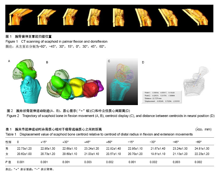
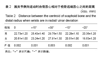
方法:纳入20名健康成人志愿者(男女各10名),均行右腕关节连续螺旋CT扫描,右手腕分别于中立位,掌屈、背伸各15°,30°,45°,60°位,桡偏10°,20°、尺偏15°,30°位进行扫描,将原始DICOM数据导入Materialise Mimics Innovation Suite 15.0软件,通过三维建模,重建桡骨远端及舟骨,并行骨块三维配准,分别测定各骨块质心,重建舟骨的运动轨迹,计算骨块间的距离。
结果与结论:腕关节屈曲及背伸或尺偏及桡偏运动时,舟骨质心相对于桡骨远端质心之间的距离是不同的,腕关节背伸60°,尺偏30°位舟骨质心相对于桡骨距离最大;背伸30°,桡偏20°,则距离最小。各位置桡骨远端质心与舟骨质心间距离男女间比较有统计学差异。实验精确测定腕舟骨的质心,为重建舟骨的运动轨迹提供了新的科学方法。

中国组织工程研究杂志出版内容重点:人工关节;骨植入物;脊柱骨折;内固定;数字化骨科;组织工程
ORCID: 0000-0002-5431-8917(张志峰)

关键词: 腕关节, 舟骨, 三维重建, 数字化, 运动轨迹

Abstract:

BACKGROUND: Scaphoid fracture is commonly seen in wrist fracture, but its nonunion rate is high. Therefore, studying trajectory of normal scaphoid bone is of great significance for selecting optimal fixation way and position.

OBJECTIVE: To study the trajectory of normal scaphoid bone, and to provide method for three-dimensional kinematics analysis.
METHODS: Twenty normal adults (n=10 in either sexes) were enrolled, and the right wrist underwent spiral CT scan in neutral, palmar flexion, dorsiflexion at 15, 30, 45 and 60 degrees, radial deviation at 10 and 20 degrees and ulnar deviation at 15 and 30 degrees, respectively. The original DICOM data were imported into Materialise Mimics Innovation Suite 15.0 to reconstruct distal radius and scaphoideum, and three-dimensional registration. The centroid of each bone was measured, and trajectory of scaphoideum was reconstructed to calculate the displacement value.
RESULTS AND CONCLUSION: In palmar flexion and dorsiflexion or radial/ulnar deviation, the distance between centroid of scaphoideum and distal radius was different. In the 60 degrees of dorsiflexion and 30 degrees of ulnar deviation, the displacement was largest. In the 30 degrees of dorsiflexion and 20 degrees of radial deviation, the displacement was smallest. The displacement value in different positions showed significant differences between sexes. In summary, the accurate determination of the centroid of scaphoid bone provides a new scientific method for the reconstruction of scaphoid movement. 

中国组织工程研究杂志出版内容重点:人工关节;骨植入物;脊柱骨折;内固定;数字化骨科;组织工程

Key words: Imaging, Three-dimensional, Scaphoid Bone, Anatomy, Tissue Engineering

中图分类号: