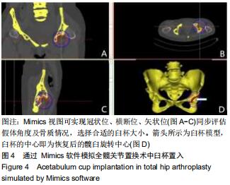
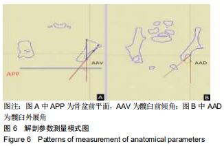
|
[1] TAMURA K, TAKAO M, HAMADA H, et al. Femoral morphology asymmetry in hip dysplasia makes radiological leg length measurement inaccurate. Bone Joint J. 2019;101-B(3):297-302.
[2] GREBER EM, PELT CE, GILILLAND JM, et al. Challenges in Total Hip Arthroplasty in the Setting of Developmental Dysplasia of the Hip. J Arthroplasty. 2017;32(9S):S38-S44.
[3] FALDINI C, NANNI M, LEONETTI D, et al. Total hip arthroplasty in developmental hip dysplasia using cementless tapered stem. Results after a minimum 10-year follow-up. Hip Int. 2011;21(4):415-420.
[4] PENG H, ZHANG G, XU C, et al. Is pseudoacetabulum an important factor determining SSTO application in total hip arthroplasty for Crowe IV hips? a retrospective cohort study. J Orthop Surg Res. 2019;14(1):201.
[5] ARGENSON JN, FLECHER X, PARRATTE S, et al. Anatomy of the dysplastic hip and consequences for total hip arthroplasty. Clin Orthop Relat Res. 2007;465:40-45.
[6] YILDIRIM T, GUCLU B, KARAGUVEN D, et al. Cementless total hip arthroplasty in developmental dysplasia of the hip with end stage osteoarthritis: 2-7 years' clinical results. Hip Int. 2015;25(5):442-446.
[7] PARK J, KIM GL, YANG KH. Anatomical landmarks for acetabular abduction in adult hips: the teardrop vs. the inferior acetabular rim. Surg Radiol Anat. 2019;41(12):1505-1511.
[8] ZHU B, SU C, HE Y, et al. Combined anteversion technique in total hip arthroplasty for Crowe IV developmental dysplasia of the hip. Hip Int. 2017;27(6):589-594.
[9] 范广,向川.髋臼假体置入角度对髋关节功能的影响[J].实用骨科杂志,2019,25(8): 740-743.
[10] KALTEIS T, HANDEL M, BÄTHIS H, et al. Imageless navigation for insertion of the acetabular component in total hip arthroplasty: is it as accurate as CT-based navigation?. J Bone Joint Surg Br. 2006;88(2):163-167.
[11] HOHMANN E, BRYANT A, TETSWORTH K. A comparison between imageless navigated and manual freehand technique acetabular cup placement in total hip arthroplasty. J Arthroplasty. 2011;26(7):1078-1082.
[12] WANG L, THORESON AR, TROUSDALE RT, et al. Two-dimensional and three-dimensional cup coverage in total hip arthroplasty with developmental dysplasia of the hip. J Biomech. 2013;46(10):1746-1751.
[13] SARIALI E, MAUPRIVEZ R, KHIAMI F, et al. Accuracy of the preoperative planning for cementless total hip arthroplasty. A randomised comparison between three-dimensional computerised planning and conventional templating. Orthop Traumatol Surg Res. 2012;98(2):151-158.
[14] XU J, LI D, MA RF, et al. Application of rapid prototyping pelvic model for patients with DDH to facilitate arthroplasty planning: a pilot study. J Arthroplasty. 2015;30(11):1963-1970.
[15] 芮敏,顾家烨,张云庆,等. 数字化三维重建技术在髋关节发育不良全髋关节置换术中的应用[J].临床骨科杂志, 2019, 22(2): 165-168.
[16] CAI Z, ZHAO Q, LI L, et al. Can computed tomography accurately measure acetabular anterversion in developmental dysplasia of the hip? verification and characterization using 3D printing technology. J Pediatr Orthop. 2018;38(4): e180-e185.
[17] 张衡,周建生. CT三维重建在成人髋关节发育不良髋臼形态研究中的进展[J].中华解剖与临床杂志,2014,19(6):519-522.
[18] 曾羿,赖欧杰,沈彬,等.计算机三维术前计划在CroweⅣ型髋关节发育不良全髋关节置换髋臼重建中的应用[J].中华骨科杂志, 2014,34(12):1212-1218.
[19] 曹力.发育性髋关节发育不良病人的全髋关节置换术:探索,挑战,求精[J].骨科,2018,9(5):337-340.
[20] AKIYAMA M, NAKASHIMA Y, FUJII M, et al. Femoral anteversion is correlated with acetabular version and coverage in Asian women with anterior and global deficient subgroups of hip dysplasia: a CT study. Skeletal Radiol. 2012; 41(11):1411-1418.
[21] FUJII M, NAKASHIMA Y, SATO T, et al. Acetabular tilt correlates with acetabular version and coverage in hip dysplasia. Clin Orthop Relat Res. 2012;470(10):2827-2835.
[22] YOSHITANI J, KABATA T, KAJINO Y, et al. Morphometric geometrical analysis to determine the centre of the acetabular component placement in Crowe type IV hips undergoing total hip arthroplasty. Bone Joint J. 2019;101-B(2):189-197.
[23] LIU RY, WANG KZ, WANG CS, et al. Evaluation of medial acetabular wall bone stock in patients with developmental dysplasia of the hip using a helical computed tomography multiplanar reconstruction technique. Acta Radiol. 2009; 50(7):791-797.
[24] 肖瑜,张福江,马信龙,等.成人髋关节发育不良不同Crowe分型的三维CT影像学特征[J].中华骨科杂志,2014,34(3):311-316.
[25] OKUZU Y, GOTO K, KAWATA T, et al. The relationship between subluxation percentage of the femoroacetabular joint and acetabular width in Asian women with developmental dysplasia of the hip. J Bone Joint Surg Am. 2017;99(7):e31.
[26] YANG Y, ZUO J, LIU T, et al. Morphological analysis of true acetabulum in hip dysplasia (Crowe Classes I-IV) Via 3-D implantation simulation. J Bone Joint Surg Am. 2017;99(17): e92.
[27] CAO L, WANG Y, ZOU S, et al. A novel positioner for accurately sitting the acetabular component: a retrospective comparative study. J Orthop Surg Res. 2019;14(1):279.
[28] SHAO P, LI Z, YANG M, et al. Impact of acetabular reaming depth on reconstruction of rotation center in primary total hip arthroplasty. BMC Musculoskelet Disord. 2018;19(1):425.
[29] KIYAMA T, NAITO M, SHINODA T, et al. Hip abductor strengths after total hip arthroplasty via the lateral and posterolateral approaches. J Arthroplasty. 2010;25(1):76-80.
[30] ASAYAMA I, CHAMNONGKICH S, SIMPSON KJ, et al. Reconstructed hip joint position and abductor muscle strength after total hip arthroplasty. J Arthroplasty. 2005;20(4):414-420.
[31] ZENG Y, LAI OJ, SHEN B, et al. Three-dimensional computerized preoperative planning of total hip arthroplasty with high-riding dislocation developmental dysplasia of the hip. Orthop Surg. 2014;6(2):95-102.
|