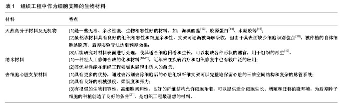
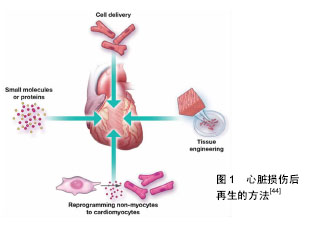
| [1] Mozaffarian D, Benjamin EJ, Go AS, Arnett DK,et al. Heart disease and stroke statistics--2015 update: a report from the American Heart Association. Circulation, United States: 2015; 131(4): e29-322.[2] 姚震,陈林. 我国心血管疾病现状与展望[J]. 海南医学,2013,24(13): 1873-1876.[3] Cahn F, Kyriakides TR. Generation of an artificial skin construct containing a non-degradable fiber mesh: a potential transcutaneous interface.Biomed Mater. 2008;3(3): 34110.[4] Nemeno-Guanzon JG, Lee S, Berg JR, et al.Trends in Tissue Engineering for Blood Vessels. J Biomed Biotechnol. 2012;2012: 956345.[5] Ling C, Li Q, Brown ME, et al. Bioengineered vocal fold mucosa for voice restoration. Sci Transl Med.2015;7(314): 314ra187.[6] Ott HC, Matthiesen TS, Goh SK, et al. Perfusion-decellularized matrix: using nature’s platform to engineer a bioartificial heart. Nature Med.2008;14(2):213-221.[7] Uzarski JS, Xia Y, Belmonte JC, et al. New strategies in kidney regeneration and tissue engineering.Curr Opin Nephrol Hypertens. 2014;23(4): 399-405.[8] Palakkan AA, Hay DC, Anil Kumar PR, et al.Liver tissue engineering and cell sources: issues and challenges.Liver Int. 2013;33(5):666-676.[9] Kador KE, Montero RB, Venugopalan P, et al. Tissue engineering the retinal ganglion cell nerve fiber layer.Biomaterials. 2013; 34(17):4242-4250.[10] Atala A.Tissue engineering of human bladder. Br Med Bull. 2011; 97:81-104.[11] Petersen TH, Calle EA, Zhao L, et al.Tissue-engineered lungs for in vivo implantation.Science. 2010;329(5991):538-541.[12] Hu XJ, Dong NG, Liu JP, et al.Status on Heart Transplantation in China.Chin Med J (Engl). 2015 Dec 5;128(23):3238-3242.[13] Li Z, Zhang M.Chitosan-alginate as scaffolding material for cartilage tissue engineering.J Biomed Mater Res A. 2005;75(2):485-493.[14] Glowacki J, Mizuno S. Collagen scaffolds for tissue engineering. Biopolymers.2008; 89(5): 338-344.[15] Zhu J, Marchant RE.Design properties of hydrogel tissue-engineering scaffolds.Expert Rev Med Devices. 2011; 8(5):607-626.[16] 徐志云,张宝仁. 组织工程心脏瓣膜的研究现状与进展[J]. 第二军医大学学报, 2003, 24(3): 233-235.[17] Hoang Thi TT, Lee JS, Lee Y, et al. Enhanced Cellular Activity in Gelatin-Poly(Ethylene Glycol) Hydrogels without Compromising Gel Stiffness.Macromol Biosci. 2016;16(3):334-340.[18] Alghazali KM, Nima ZA, Hamzah RN, et al.Bone-tissue engineering: complex tunable structural and biological responses to injury, drug delivery, and cell-based therapies. Drug Metab Rev. 2015;47(4):431-454.[19] Karunaratne A, Xi L, Bentley L, et al. Multiscale alterations in bone matrix quality increased fragility in steroid induced osteoporosis. Bone. 2016;84:15-24.[20] Kim SY, Hwang JY, Shin US. Preparation of nano/macroporous polycaprolactone microspheres for an injectable cell delivery system using room temperature ionic liquid and camphene.J Colloid Interface Sci. 2016;465:18-25[21] 王圣,李温斌.去细胞组织工程心脏瓣膜研究现状与展望[J]. 中国医疗器械信息,2009,15(2):14-17.[22] Bai R,Liu HL.Heart decellularized matrix application in cardiac tissue engineering.Jiefangjun Yixueyuan Xuebao.2015;7:1-4.[23] Rieder E, Kasimir MT, Silberhumer G, et al. Decellularization protocols of porcine heart valves differ importantly in efficiency of cell removal and susceptibility of the matrix to recellularization with human vascular cells. J Thorac Cardiovasc Surg. 2004; 127(2):399-405.[24] Hussein KH, Park KM, Kang KS, et al.Biocompatibility evaluation of tissue-engineered decellularized scaffolds for biomedical application.Mater Sci Eng C Mater Biol Appl. 2016;67:766-778.[25] Papalamprou A, Griffiths LG. Cardiac Extracellular Matrix Scaffold Generated Using Sarcomeric Disassembly and Antigen Removal.Ann Biomed Eng. 2016;44(4):1047-1060.[26] Papalamprou A, Chang CW, Vapniarsky N,et al. Xenogeneic cardiac extracellular matrix scaffolds with or without seeded mesenchymal stem cells exhibit distinct in vivo immunosuppressive and regenerative properties. Acta Biomaterialia.2016;45:155-168.[27] Li Q, Uygun BE, Geerts S, et al.Proteomic Analysis of Naturally-Sourced Biological Scaffolds. Biomaterials.2015;75: 37-46.[28] HERRMANN F E M, LEHNER A, HOLLWECK T et al. In vitro biological and mechanical evaluation of various scaffold materials for myocardial tissue engineering.J Biomed Mater Res A. 2014; 102(4):958-966.[29] 肖统光. 细胞外基质来源支架在软骨组织工程中的应用[J]. 中国组织工程研究,2016,20(38):5737-5744.[30] 杨立信.骨髓间质干细胞构建组织工程心脏瓣膜的实验研究[J]. 第二军医大学.2004.[31] 黄胜兰,吴敬波. 心肌细胞增殖的研究进展[J]. 现代临床医学, 2011, 37(6):9-11.[32] Matsuyama D, Kawahara K.Proliferation of neonatal cardiomyocytes by connexin43 knockdown via synergistic inactivation of p38 MAPK and increased expression of FGF1. Basic Res Cardiol. 2009;104(6):631-642.[33] Ebelt H, Zhang Y, Köhler K, et al. Directed expression of dominant-negative p73 enables proliferation of cardiomyocytes in mice.J Mol Cell Cardiol. 2008;45(3):411-419.[34] Puente BN, Kimura W, Muralidhar SA, et al.The oxygen-rich postnatal environment induces cardiomyocyte cell-cycle arrest through DNA damage response. Cell.2014;157(3): 565-579.[35] Chamberlain G, Fox J, Ashton B,et al.Concise Review: Mesenchymal Stem Cells: Their Phenotype, Differentiation Capacity, Immunological Features, and Potential for Homing. Stem Cells.2007; 25(11):2739-2749.[36] Stubbs SL, Crook JM, Morrison WA, et al.Toward Clinical Application of Stem Cells for Cardiac RegenerationHeart Lung Circ. 2011;20(3):173-179.[37] Sun L, Cui M, Wang Z,et al. Mesenchymal stem cells modified with angiopoietin-1 improve remodeling in a rat model of acute myocardial infarction.Biochem Biophys Res Commun. 2007; 357(3):779-784.[38] de Macedo Braga LM, Lacchini S, Schaan BD, et al.In situ delivery of bone marrow cells and mesenchymal stem cells improves cardiovascular function in hypertensive rats submitted to myocardial infarction.J Biomed Sci. 2008;15(3):365-374.[39] 王福科,刘流,李俊男,等.间充质干细胞在组织工程中的应用进展[J].中国组织工程研究与临床康复,2010,14(36): 6800-6804.[40] Crisan M, Yap S, Casteilla L, et al.A Perivascular Origin for Mesenchymal Stem Cells in Multiple Human Organs. Cell Stem Cell.2008;3(3): 301-313.[41] Kocher AA, Schuster MD, Szabolcs MJ, et al. Neovascularization of ischemic myocardium by human bone-marrow-derived angioblasts prevents cardiomyocyte apoptosis, reduces remodeling and improves cardiac function.Nature Med. 2001;7(4): 430-436.[42] Antonitsis P, Ioannidou-Papagiannaki E, Kaidoglou A, et al. In vitro cardiomyogenic differentiation of adult human bone marrow mesenchymal stem cells. The role of 5-azacytidine.Interact Cardiovasc Thorac Surg. 2007;6(5):593-597.[43] Carvalho PH, Daibert AP, Monteiro BS, et al. Differentiation of adipose tissue-derived mesenchymal stem cells into cardiomyocytes. Arq Bras Cardiol. 2013;100(1):82-89.[44] Garbern JC, Lee RT. Cardiac stem cell therapy and the promise of heart regeneration.Cell Stem Cell. 2013;12(6):689-698.[45] Kang H, Lu S, Peng J, et al.In vivo construction of tissue-engineered cartilage using adipose-derived stem cells and bioreactor technology.Cell Tissue Bank. 2015;16(1):123-133.[46] Guyette JP, Charest JM, Mills RW, et al. Bioengineering Human Myocardium on Native Extracellular Matrix.Circ Res. 2016;118(1): 56-72.[47] Weymann A, Patil NP, Sabashnikov A, et al.Bioartificial heart: a human-sized porcine model--the way ahead.PLoS One. 2014 ; 9(11):e111591.[48] Tao ZW, Mohamed M, Hogan M, et al.Establishing the Framework for Fabrication of a Bioartificial Heart.ASAIO J. 2015;61(4): 429-436.[49] Hirt MN, Boeddinghaus J, Mitchell A, et al.Functional improvement and maturation of rat and human engineered heart tissue by chronic electrical stimulation.J Mol Cell Cardiol. 2014;74: 151-161.[50] He Q, Johnston J, Zeitlinger J.ChIP-nexus enables improved detection of in vivo transcription factor binding footprints. HHS Public Access.2015;33(4): 395–401.[51] 温华知. 腺病毒介导的Sema3A基因对大鼠心肌梗死后交感神经重构和电重构的影响[D]. 武汉大学, 2010.[52] Alteköester AK, Harvey RP.Bioengineered FSTL1 Patches Restore Cardiac Function Following Myocardial Infarction.Trends Mol Med. 2015;21(12):731-733.[53] Rimmbach C, Jung JJ, David R.Generation of Murine Cardiac Pacemaker Cell Aggregates Based on ES-Cell-Programming in Combination with Myh6-Promoter-Selection.J Vis Exp. 2015;(96): e52465. |
.jpg)
.jpg)