[1] GRITSCH S, BATCHELOR TT, GONZALEZ CASTRO LN. Diagnostic, therapeutic, and prognostic implications of the 2021 World Health Organization classification of tumors of the central nervous system. Cancer. 2022;128(1):47-58.
[2] GONDAR R, PATET G, SCHALLER K, et al. Meningiomas and Cognitive Impairment after Treatment: A Systematic and Narrative Review. Cancers (Basel). 2021;13(8): 1846.
[3] CORNIOLA MV, MELING TR. Management of Recurrent Meningiomas: State of the Art and Perspectives. Cancers (Basel). 2022;14(16):3995.
[4] LI X, HE S, MA B. Autophagy and autophagy-related proteins in cancer. Mol Cancer. 2020;19(1):12.
[5] ANTUNES F, ERUSTES AG, COSTA AJ, et al. Autophagy and intermittent fasting: the connection for cancer therapy? Clinics (Sao Paulo). 2018;73(suppl 1):e814s.
[6] USMAN RM, RAZZAQ F, AKBAR A, et al. Role and mechanism of autophagy-regulating factors in tumorigenesis and drug resistance. Asia Pac J Clin Oncol. 2021;17(3):193-208.
[7] FENG X, ZHANG H, MENG L, et al. Hypoxia-induced acetylation of PAK1 enhances autophagy and promotes brain tumorigenesis via phosphorylating ATG5. Autophagy. 2021;17(3):723-742.
[8] WHITE E. The role for autophagy in cancer. J Clin Invest. 2015;125(1):42-46.
[9] ZAMAME RAMIREZ JA, ROMAGNOLI GG, KANENO R. Inhibiting autophagy to prevent drug resistance and improve anti-tumor therapy. Life Sci. 2021;265:118745.
[10] LIU W, MENG Y, ZONG C, et al. Autophagy and Tumorigenesis. Adv Exp Med Biol. 2020;1207:275-299.
[11] WANG Y, DU J, WU X, et al. Crosstalk between autophagy and microbiota in cancer progression. Mol Cancer. 2021;20(1):163.
[12] PARK S, CHA YJ, SUH SH, et al. Risk group-adapted adjuvant radiotherapy for WHO grade I and II skull base meningioma. J Cancer Res Clin Oncol. 2019;145(5): 1351-1360.
[13] MAGGIO I, FRANCESCHI E, TOSONI A, et al. Meningioma: not always a benign tumor. A review of advances in the treatment of meningiomas. CNS Oncol. 2021; 10(2):CNS72.
[14] HORBINSKI C, BERGER T, PACKER RJ, et al. Clinical implications of the 2021 edition of the WHO classification of central nervous system tumours. Nat Rev Neurol. 2022;18(9):515-529.
[15] PREUSSER M, BRASTIANOS PK, MAWRIN C. Advances in meningioma genetics: novel therapeutic opportunities. Nat Rev Neurol. 2018;14(2):106-115.
[16] KLIONSKY DJ, PETRONI G, AMARAVADI RK, et al. Autophagy in major human diseases. EMBO J. 2021;40(19):e108863.
[17] CHEN X, YU C, KANG R, et al. Cellular degradation systems in ferroptosis. Cell Death Differ. 2021;28(4):1135-1148.
[18] LI W, HE P, HUANG Y, et al. Selective autophagy of intracellular organelles: recent research advances. Theranostics. 2021;11(1):222-256.
[19] CAO W, LI J, YANG K, et al. An overview of autophagy: Mechanism, regulation and research progress. Bull Cancer. 2021;108(3):304-322.
[20] ONORATI AV, DYCZYNSKI M, OJHA R, et al. Targeting autophagy in cancer. Cancer. 2018;124(16):3307-3318.
[21] YANG K, NIU L, BAI Y, et al. Glioblastoma: Targeting the autophagy in tumorigenesis. Brain Res Bull. 2019;153:334-340.
[22] YAMAZAKI T, BRAVO-SAN PEDRO JM, GALLUZZI L, et al. Autophagy in the cancer-immunity dialogue. Adv Drug Deliv Rev. 2021;169:40-50.
[23] XU Z, HAN X, OU D, et al. Targeting PI3K/AKT/mTOR-mediated autophagy for tumor therapy. Appl Microbiol Biotechnol. 2020;104(2):575-587.
[24] SMITH AG, MACLEOD KF. Autophagy, cancer stem cells and drug resistance. J Pathol. 2019;247(5):708-718.
[25] COCCO S, LEONE A, PIEZZO M, et al. Targeting Autophagy in Breast Cancer. Int J Mol Sci. 2020 22;21(21):7836.
[26] 曲宝清,米蕊芳,杜江,等. 脑胶质瘤超微结构及组织形态观察39例[J].中国临床医生杂志,2019,47(5):63-65.
[27] KOUSTAS E, SARANTIS P, THEOHARIS S, et al. Autophagy-related Proteins as a Prognostic Factor of Patients With Colorectal Cancer. Am J Clin Oncol. 2019; 42(10):767-776.
[28] ROY BC, AHMED I, RAMALINGAM S, et al. Co-localization of autophagy-related protein p62 with cancer stem cell marker dclk1 may hamper dclk1’s elimination during colon cancer development and progression. Oncotarget. 2019;10(24):2340-2354.
[29] TOTON E, LISIAK N, SAWICKA P, et al. Beclin-1 and its role as a target for anticancer therapy. J Physiol Pharmacol. 2014;65(4):459-467.
[30] HAMURCU Z, DELIBAŞI N, GEÇENE S, et al. Targeting LC3 and Beclin-1 autophagy genes suppresses proliferation, survival, migration and invasion by inhibition of Cyclin-D1 and uPAR/Integrin β1/ Src signaling in triple negative breast cancer cells. J Cancer Res Clin Oncol. 2018;144(3):415-430.
[31] HU Y, LI X, XUE W, et al. TP53INP2-related basal autophagy is involved in the growth and malignant progression in human liposarcoma cells. Biomed Pharmacother. 2017;88:562-568.
[32] YING H, QU D, LIU C, et al. Chemoresistance is associated with Beclin-1 and PTEN expression in epithelial ovarian cancers. Oncol Lett. 2015;9(4):1759-1763.
[33] WANG L, WANG J, XIONG H, et al. Co-targeting hexokinase 2-mediated Warburg effect and ULK1-dependent autophagy suppresses tumor growth of PTEN- and TP53-deficiency-driven castration-resistant prostate cancer. EBioMedicine. 2016; 7:50-61.
[34] ABDELZAHER E, EL-GENDI SM, YEHYA A, et al. Recurrence of benign meningiomas: predictive value of proliferative index, BCL2, p53, hormonal receptors and HER2 expression. Br J Neurosurg. 2011;25(6):707-713.
[35] DOMINGUES PH, SOUSA P, OTERO Á, et al. Proposal for a new risk stratification classification for meningioma based on patient age, WHO tumor grade, size, localization, and karyotype. Neuro Oncol. 2014;16(5):735-747.
[36] FOLKERTS H, HILGENDORF S, VELLENGA E, et al. The multifaceted role of autophagy in cancer and the microenvironment. Med Res Rev. 2019;39(2):517-560.
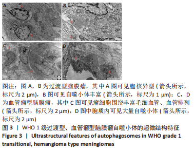
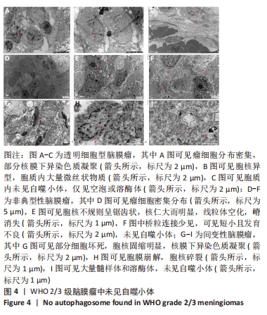
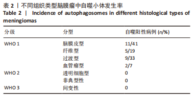
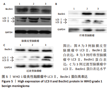
|