中国组织工程研究 ›› 2023, Vol. 27 ›› Issue (33): 5298-5303.doi: 10.12307/2023.514

• 干细胞基础实验 basic experiments of stem cells • 上一篇    下一篇

Ngn2调节Nrf2/HO-1对脑缺血模型大鼠脑微结构、角质细胞活性的影响

王明盛1,崔焕喜1,崔红凯1,裴观辉1,李道广1,闫海清2,张  平2   

  1. 新乡医学院第一附属医院,1神经介入科,2神经内科,河南省卫辉市  453100
  • 收稿日期:2022-06-18 接受日期:2022-08-19 出版日期:2023-11-28 发布日期:2023-03-30
  • 通讯作者: 张平,博士,主任医师,新乡医学院第一附属医院神经内科,河南省卫辉市 453100
  • 作者简介:王明盛,男,1974年生,河南省焦作市人,汉族,2005年华中科技大学毕业,硕士,副主任医师,主要从事脑血管病综合治疗的研究。
  • 基金资助:
    河南省医学科技公关计划省部共建项目(SB201901061),项目负责人:张平;河南省医学科技公关计划联合共建项目(LHGJ20200488),项目负责人:闫海清

Ngn2 effects on brain microstructure and keratinocyte activity in cerebral ischemia rats via regulating Nrf2/HO-1

Wang Mingsheng1, Cui Huanxi1, Cui Hongkai1, Pei Guanhui1, Li Daoguang1, Yan Haiqing2, Zhang Ping2   

  1. 1Department of Neurointervention, 2Department of Neurology, The First Affiliated Hospital of Xinxiang Medical University, Weihui 453100, Henan Province, China
  • Received:2022-06-18 Accepted:2022-08-19 Online:2023-11-28 Published:2023-03-30
  • Contact: Zhang Ping, MD, Chief physician, Department of Neurology, The First Affiliated Hospital of Xinxiang Medical University, Weihui 453100, Henan Province, China
  • About author:Wang Mingsheng, Master, Associate chief physician, Department of Neurointervention, The First Affiliated Hospital of Xinxiang Medical University, Weihui 453100, Henan Province, China
  • Supported by:
    Henan Province Medical Science and Technology Public Relations Plan - Provincial and Ministry Co-Construction Project, No. SB201901061 (to ZP); Henan Province Medical Science and Technology Public Relations Plan - Joint Construction Project, No. LHGJ20200488 (to YHQ)

摘要:


文题释义:

脑缺血:指由于多种原因导致的局部或多部位的供血不足性疾病。其主要病因与脑动脉狭窄或闭塞、脑动脉栓塞、血流动力学因素、血液学因素有关,主要临床症状包括头痛、呕吐、眩晕、偏瘫、感觉障碍,可导致癫痫、面瘫、语言障碍等并发症。目前主要通过溶栓、手术、药物等多方式治疗,预后与疾病类型有关。
脑微结构:脑结构可塑性改变是脑损伤后功能恢复的基础。神经系统中细胞与细胞之间的信号依靠突触结构传递,突触是神经递质和受体作用的功能单位,其结构的完整性是神经元正常生理功能的物质基础。突触超微结构变化也是衡量脑结构可塑性的重要方面。

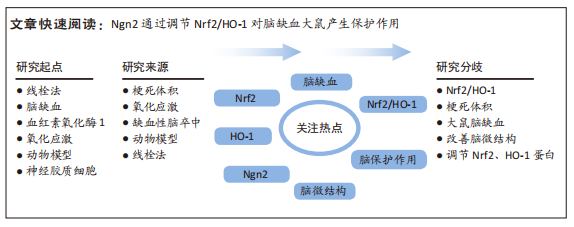
背景:研究显示,血红素氧化酶1(heme oxidase-1,HO-1)在脑缺血再灌注损伤中具有重要作用;核因子E2相关因子2(nuclear factor-erythroid2-related factor2,Nrf2)能减轻脑缺血再灌注损伤,其作用是通过调控下游抗氧化蛋白实现的;推测Nrf2/HO-1在脑部疾病中均有一定的调控作用。
目的:探究神经源素2(neurogenin 2,Ngn2)通过调节Nrf2/HO-1对脑缺血大鼠脑微结构、角质细胞活性的影响。
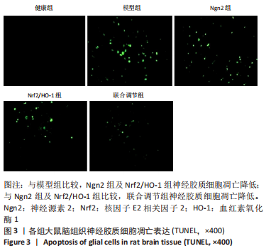
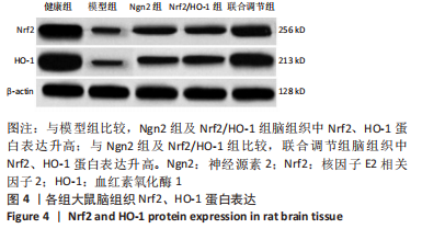
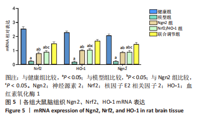
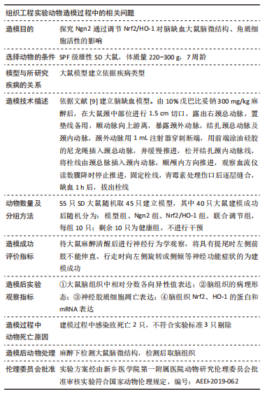
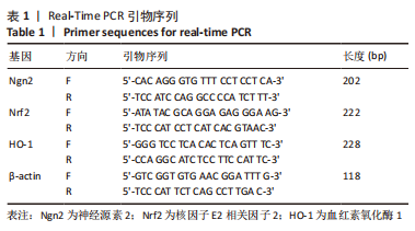
方法:SPF级雄性SD大鼠55只,随机取10只为健康组不进行干预;其余大鼠建立脑缺血模型,将建模成功的40只大鼠随机分为4组:其中模型组大鼠脑内注射生理盐水;Ngn2组大鼠脑内注射Ngn2 10 mg/kg;Nrf2/HO-1组脑内注射HO-1及Nrf2激动剂莱菔硫烷各10 mg/kg;联合调节组脑内注射Ngn2并联合Nrf2/HO-1组用药,均连续给药3 d。分数各向异性图像观察脑微结构,苏木精-伊红染色观察脑组织的病理形态,TUNEL法检测神经胶质细胞凋亡,免疫印迹和PCR分别检测Nrf2、HO-1的蛋白及mRNA表达。

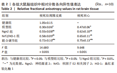
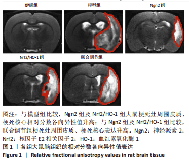
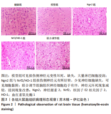
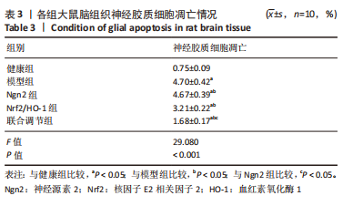
结果与结论:①与健康组比较,模型组大鼠脑组织中梗死灶周围皮质、梗死核心相对分数各向异性值表达较低(P < 0.05);与模型组比较,Ngn2组及Nrf2/HO-1组上述表达升高(P < 0.05);联合调节组上述表达高于Ngn2组及Nrf2/HO-1组(P < 0.05);②模型组大鼠大量损伤神经元,细胞稀疏,排列紊乱,大量浸润;Ngn2组与Nrf2/HO-1组损伤侧神经元好转,仍见神经细胞缺失紊乱及细胞黏附;联合调节组脑组织神经细胞坏死减轻,浸润改善;③与健康组比较,模型组大鼠神经胶质细胞凋亡较高(P < 0.05);与模型组比较,Ngn2组及Nrf2/HO-1组神经胶质细胞凋亡降低(P < 0.05);联合调节组神经胶质细胞凋亡低于Ngn2组及Nrf2/HO-1组(P < 0.05);④与健康组比较,模型组大鼠脑组织Ngn2 mRNA及Nrf2、HO-1的蛋白和mRNA表达较低(P < 0.05);与模型组比较,Ngn2组、Nrf2/HO-1组Ngn2 mRNA及Nrf2、HO-1的蛋白和mRNA表达升高(P < 0.05);联合调节组Ngn2 mRNA及Nrf2、HO-1的蛋白和mRNA表达高于Ngn2组及Nrf2/HO-1组(P < 0.05);⑤结果说明,Ngn2通过调节Nrf2/HO-1对脑缺血大鼠产生保护作用,其机制可能与改善脑微结构、角质细胞活性以及增强Nrf2、HO-1表达等具有一定相关性。

https://orcid.org/0000-0001-6610-0141(王明盛)

中国组织工程研究杂志出版内容重点:干细胞;骨髓干细胞;造血干细胞;脂肪干细胞;肿瘤干细胞;胚胎干细胞;脐带脐血干细胞;干细胞诱导;干细胞分化;组织工程

关键词: 脑缺血, Ngn2基因, 核因子E2相关因子2, 血红素氧化酶1, Nrf2/HO-1, 脑微结构, 角质细胞活性

Abstract: BACKGROUND: Heme oxidase-1 (HO-1) plays an important role in cerebral ischemia-reperfusion injury. Nuclear factor-erythroid 2-related factor 2 (Nrf2) can reduce cerebral ischemia-reperfusion injury via regulating downstream antioxidant proteins. Therefore, it is speculated that Nrf2/HO-1 has a certain regulatory role in brain diseases.
OBJECTIVE: To explore the effects of Neurogenin 2 (Ngn2) on brain microstructure and keratinocyte activity in cerebral ischemia rats by regulating Nrf2/HO-1.
METHODS: Fifty-five SPF male Sprague-Dawley rats were selected, 10 of which were randomly selected as healthy group and the remaining rats were used to establish animal models of cerebral ischemia. After modeling, 40 model rats were randomly divided into 4 groups: rats in model group were intracerebrally injected with normal saline; rats in Ngn2 group were intracerebrally injected with Ngn2 10 mg/kg; rats in Nrf2/HO-1 group was intracerebrally injected with Ho-1 and Nrf2 agonist sulforaphane 10 mg/kg; and rats in combined group received intracerebral injection of Ngn2 combined with Nrf2/HO-1 10 mg/kg. Administration in each group lasted for 3 consecutive days. Fractional anisotropy imaging was used to observe the brain microstructure. Hematoxylin-eosin staining was used to observe the pathological morphology of brain tissue. TUNEL method was used to detect glial cell apoptosis. Western blot and PCR were used to detect Nrf2 and HO-1 protein and mRNA expressions, respectively.
RESULTS AND CONCLUSION: (1) Compared with the healthy group, the relative fraction anisotropy values of the peri-infarct cortex and infarct core were lower in the model group (P < 0.05). Compared with the model group, the relative fraction anisotropy values of peri-infarct cortex and infarct core were increased in the Ngn2 group and Nrf2/HO-1 group (P < 0.05). Compared with the Ngn2 group and Nrf2/HO-1 group, the relative fraction anisotropy values of peri-infarct cortex and infarct core were increased in the combined group (P < 0.05). (2) In the model group, a large number of neurons were damaged, and the cells were sparse, disordered and infiltrated. In the Ngn2 group and Nrf2/HO-1 group, the neurons on the injured side were improved, and the loss, disorder, and adhesion of nerve cells were still observed. In the combined group, the necrosis of nerve cells was reduced and cell infiltration was improved. (3) Compared with the healthy group, the apoptosis of glial cells was higher in the model group (P < 0.05). Compared with the model group, the apoptosis of glial cells was decreased in the Ngn2 group and Nrf2/HO-1 group (P < 0.05). Compared with the Ngn2 group and Nrf2/HO-1 group, the apoptosis of glial cells was decreased in the combined group (P < 0.05). (4) Compared with the healthy group, the expressions of Ngn2 mRNA and Nrf2 and HO-1 at protein and mRNA levels were lower in the model group (P < 0.05). Compared with the model group, the expressions of Ngn2 mRNA and Nrf2 and HO-1 at protein and mRNA levels were increased in the Ngn2 group and Nrf2/HO-1 group (P < 0.05). Compared with the Ngn2 group and Nrf2/HO-1 group, the expressions of Ngn2 mRNA and Nrf2 and HO-1 at protein and mRNA levels were increased in the combined group (P < 0.05). (5) To conclude, Ngn2 can protect rats from cerebral ischemia by regulating Nrf2/HO-1, and the mechanism may be related to improving brain microstructure, keratinocyte activity and expression of Nrf2 and HO-1.

Key words: cerebral ischemia, Ngn2 gene, nuclear factor-erythroid 2-related factor 2, heme oxidase-1, Nrf2/HO-1, brain microstructure, keratinocyte activity

中图分类号: