[1] LIU C, XUE J, LIU J, et al. Is there a correlation between upper lumbar disc herniation and multifidus muscle degeneration? A retrospective study of MRI morphology. BMC Musculoskelet Disord. 2021;22(1):92.
[2] XU W, YANG B, LAI X, et al. Comparison of microendoscopic discectomy and percutaneous transforaminal endoscopic discectomy for upper lumbar disc herniation: A protocol for a systematic review and meta-analysis. Medicine (Baltimore). 2021;100(46):e27914.
[3] HUET T, COHEN-SOLAL M, LAREDO JD, et al. Lumbar spinal stenosis and disc alterations affect the upper lumbar spine in adults with achondroplasia. Sci Rep. 2020;10(1):4699.
[4] PARK JH, CHUNG SG, KIM K. Electrodiagnostic characteristics of upper lumbar stenosis: Discrepancy between neurological and structural levels. Muscle Nerve. 2020;61(5):580-586.
[5] KARAASLAN B, ASLAN A, BÖRCEK AÖ, et al. Clinical and surgical outcomes of upper lumbar disc herniations: a retrospective study. Turk J Med Sci. 2017;47(4):1157-1160.
[6] LEE DS, PARK KS, PARK MS. The Comparative Analysis of Clinical Characteristics and Surgical Results between the Upper and Lower Lumbar Disc Herniations. J Korean Neurosurg Soc. 2013;54(5):379-383.
[7] LI Z, ZHANG C, CHEN W, et al. Percutaneous Endoscopic Transforaminal Discectomy versus Conventional Open Lumbar Discectomy for Upper Lumbar Disc Herniation: A Comparative Cohort Study. Biomed Res Int. 2020;2020:1852070.
[8] YÜCE I, KAHYAOĞLU O, MERTAN P, et al. Analysis of clinical characteristics and surgical results of upper lumbar disc herniations. Neurochirurgie. 2019;65(4):158-163.
[9] SHIN MH, BAE JS, CHO HL, et al. Extradiscal Epiduroscopic Percutaneous Endoscopic Discectomy for Upper Lumbar Disc Herniation A Technical Note. Clin Spine Surg. 2019;32(3):98-103.
[10] HERRING E, KASLIWAL MK. Commentary: Novel Transdural Epiarachnoid Approach for Large Central Disk Herniation in Upper Lumbar Spine. Oper Neurosurg (Hagerstown). 2022;22(2):e111-e112.
[11] VIRK S, VAISHNAV AS, SHEHA E, et al. Combining Expandable Interbody Cage Technology With a Minimally Invasive Technique to Harvest Iliac Crest Autograft Bone to Optimize Fusion Outcomes in Minimally Invasive Transforaminal Lumbar Interbody Fusion Surgery. Clin Spine Surg. 2021;34(9):E522-E530.
[12] ZHAO J, ZHANG S, LI X, et al. Comparison of Minimally Invasive and Open Transforaminal Lumbar Interbody Fusion for Lumbar Disc Herniation: A Retrospective Cohort Study. Med Sci Monit. 2018;24: 8693-8698.
[13] XUE J, SONG Y, LIU H, et al. Minimally invasive versus open transforaminal lumbar interbody fusion for single segmental lumbar disc herniation: A meta-analysis. J Back Musculoskelet Rehabil. 2022; 35(3):505-516.
[14] XIAO C, YIN W, ZHAO K, et al. Early Clinical Efficacy of Endo-TLIF in the Treatment of Lumbar Disc Herniation [published online ahead of print, 2022 Apr 25]. Frühe klinische Wirksamkeit von Endo-TLIF bei der Behandlung von lumbalen Bandscheibenvorfällen - Bandscheibenvorfall und lumbale Instabilität: eine vergleichende Studie mit der traditionellen Methode der Behandlung [published online ahead of print, 2022 Apr 25]. Z Orthop Unfall. 2022;10.1055/a-1795-4038.
[15] YANG Y, YAN X, LI W, et al. Long-Term Clinical Outcomes and Pain Assessment after Posterior Lumbar Interbody Fusion for Recurrent Lumbar Disc Herniation. Orthop Surg. 2020;12(3):907-916.
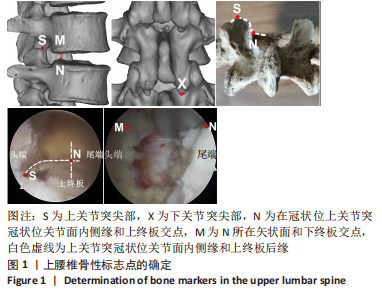
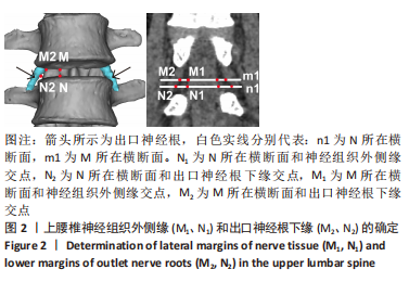
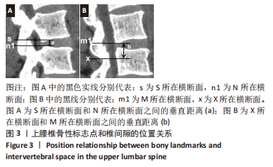
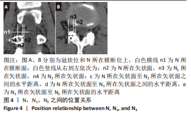
[16] 杨福生,王文,康宁超,等 .腰椎脊柱内镜微创手术中区域定位原则[J]. 中国疼痛医学杂志,2017,23(8):619-621.
[17] 王建业,刘鑫,田霖,等.构建腰椎三维模型测量腰椎管减压术区神经组织及骨性结构的位置关系[J].中国组织工程研究,2023,27(4): 539-546.
[18] 王建业,刘鑫,任佳彬,等.单侧双通道脊柱内镜技术对侧入路治疗上腰椎椎间盘突出症的影像学研究及临床应用[J].中国修复重建外科杂志,2022,36(10):1213-1220.
[19] 毕经纬,任佳彬,刘鑫,等.单侧双通道内镜的上段腰椎影像解剖测量[J].中国矫形外科杂志,2022,30(14):1293-1298.
[20] ERDOĞAN U. The Results of Using a Transforaminal Lumbar Interbody Fusion Cage at the Upper Lumbar Level. Cureus. 2021;13(6):e15496.
[21] KANG J, CHANG Z, HUANG W, et al. The posterior approach operation to treat thoracolumbar disc herniation: A minimal 2-year follow-up study. Medicine (Baltimore). 2018;97(16):e0458.
[22] WU J, ZHANG C, ZHENG W, et al. Analysis of the Characteristics and Clinical Outcomes of Percutaneous Endoscopic Lumbar Discectomy for Upper Lumbar Disc Herniation. World Neurosurg. 2016;92:142-147.
[23] BAE J, LEE SH, SHIN SH, et al. Radiological analysis of upper lumbar disc herniation and spinopelvic sagittal alignment. Eur Spine J. 2016; 25(5):1382-1388.
[24] NISHIKAWA H, FUJIMOTO M, TANIOKA S, et al. Novel Transdural Epiarachnoid Approach for Large Central Disk Herniation in Upper Lumbar Spine. Oper Neurosurg (Hagerstown). 2022;22(1):e58-e61.
[25] JHA RT, SYED HR, CATALINO M, et al. Contralateral Approach for Minimally Invasive Treatment of Upper Lumbar Intervertebral Disc Herniation: Technical Note and Case Series. World Neurosurg. 2017; 100:583-589.
[26] SON S, LEE SG, KIM WK, et al. Advantages of a Microsurgical Translaminar Approach (Keyhole Laminotomy) for Upper Lumbar Disc Herniation. World Neurosurg. 2018;119:e16-e22.
[27] MCCARTHY M, SWIATEK PR, ROUMELIOTIS AG, et al. Comparison of Lumbar Fusion With and Without Interbody Fusion for Lumbar Stenosis Using Patient-Reported Outcomes Measurement Information System (PROMIS) Computer Adaptive Testing (CAT). Cureus. 2022; 14(3):e23467.
[28] YUAN C, WANG J, ZHOU Y, et al. Endoscopic lumbar discectomy and minimally invasive lumbar interbody fusion: a contrastive review. Wideochir Inne Tech Maloinwazyjne. 2018;13(4):429-434.
[29] LIU C, ZHOU Y. Comparison Between Percutaneous Endoscopic Lumbar Discectomy and Minimally Invasive Transforaminal Lumbar Interbody Fusion for Lumbar Disc Herniation with Biradicular Symptoms. World Neurosurg. 2018;120:e72-e79.
[30] CASTILLO H, CHINTAPALLI RTV, BOYAJIAN HH, et al. Lumbar discectomy is associated with higher rates of lumbar fusion. Spine J. 2019;19(3): 487-492.
[31] 李瑞,孙兆忠,房清敏,等.椎间孔镜TESSYS技术上关节突磨削程度对腰椎稳定性的影响[J].中国矫形外科杂志,2018,26(10):898-903.
[32] SORIANO-SÁNCHEZ JA, QUILLO-OLVERA J, SORIANO-SOLIS S, et al. Microscopy-assisted interspinous tubular approach for lumbar spinal stenosis. J Spine Surg. 2017;3(1):64-70.
[33] 吐尔洪江·阿布都热西提,孟祥玉,买合木提·亚库甫,等.椎间孔镜下腰椎间盘髓核摘除治疗腰椎间盘突出症的生物力学优势[J].中国组织工程研究,2020,24(36):5768-5773.
[34] 余洋,谢一舟,石银,等.三维有限元法分析腰椎不同尺寸关节突成形后相关节段的生物力学特征[J].中国组织工程研究,2021, 25(33):5288-5293.
[35] 朱爱国,张烽,朱建炜,等.腰骶丛神经根的应用解剖及临床意义[J].中国组织工程研究,2019,23(4):573-577.
[36] ZHANG L, YANG J, HAI Y, et al. Relationship of the Exiting Nerve Root and Superior Articular Process in Kambin’s Triangle: Assessment of Lumbar Anatomy Using Cadavers and Computed Tomography Imaging. World Neurosurg. 2020;137:e336-e342.
[37] CAN H, UNAL TC, DOLAS I, et al. Comprehensive Anatomic and Morphometric Analyses of Triangular Working Zone for Transforaminal Endoscopic Approach in Lumbar Spine: A Fresh Cadaveric Study. World Neurosurg. 2020;138:e486-e491.
[38] YAMADA K, NAGAHAMA K, ABE Y, et al. Morphological analysis of Kambin’s triangle using 3D CT/MRI fusion imaging of lumbar nerve root created automatically with artificial intelligence. Eur Spine J. 2021; 30(8):2191-2199.
[39] ZHAO X, ZHAO J, GUAN J, et al. Measurement of the nerve root of the lower lumbar region using digital images. Medicine (Baltimore). 2018;97(8):e9848.
[40] ZHANG KH, ZHANG WH, XU BS, et al. CT-based Morphometric Analysis of Approach of Percutaneous Transforaminal Endoscopic Lumbar Interbody Fusion. Orthop Surg. 2019;11(2):212-220. |
 综上所述,对于上腰椎椎间盘突出症常规开放手术,OSE更容易实现潜行减压,避免了骨性结构的过度破坏,同时12°内镜可制备良好的终板植骨床,提高了术后融合率。通过三维重建技术、影像学解剖相结合,测量相关数据为OSE技术的临床手术提供指导,使手术过程更加精准、安全。但此次研究也存在不足之处:纳入研究的上腰椎样本量较少;在广泛群体中存在一定的局限性,多适用在年龄群体偏大的患者;下一步会扩大样本量,深入研究广泛群体以此来指导临床工作。