[1] VASQUEZ C, YANG A, YOUSSEF AS. Foramen Magnum Meningioma: Far Lateral Approach. J Neurol Surg B Skull Base. 2019;80(Suppl 4): S363-S364.
[2] Beer-Furlan A, Vellutini EA, Balsalobre L, et al. Endoscopic Endonasal Approach to Ventral Posterior Fossa Meningiomas: From Case Selection to Surgical Management. Neurosurg Clin N Am. 2015; 26(3):413-426.
[3] AMELOT A, TERRIER LM, LOT G. Craniovertebral Junction Transoral Approach: Predictive Factors of Complications. World Neurosurg. 2018; 110:568-574.
[4] Heros RC. Lateral suboccipital approach for vertebral and vertebrobasilar artery lesions. J Neurosurg. 1986;64(4):559-562.
[5] NANDA A, VINCENT DA, VANNEMREDDY PS, et al. Far-lateral approach to intradural lesions of the foramen magnum without resection of the occipital condyle. J Neurosurg. 2002;96(2):302-309.
[6] BASSIOUNI H , NTOUKAS V, ASGARI S, et al. Foramen magnum meningiomas: clinical outcome after microsurgical resection via a posterolateral suboccipital retrocondylar approach. Neurosurgery. 2006;59(6):1177-1185.
[7] BOULTON MR , CUSIMANO MD. Foramen magnum meningiomas: concepts, classifications, and nuances. Neurosurg Focus. 2003;14(6): e10.
[8] BOLZ S, GAPERT R, HARTWIG S, et al. Evaluation of foramen magnum sexual dimorphism in a modern documented German population using post-mortem computed tomography. Forensic Imaging. 2020; 21:200352.
[9] TONEVA D, NIKOLOVA S, HARIZANOV S, et al. Sex estimation by size and shape of foramen magnum based on CT imaging. Leg Med (Tokyo). 2018;35:50-60.
[10] TEIXEIRA WR. Sex identification utilizing the size of the foramen magnum. Am J Forensic Med Pathol. 1982;3(3):203-206.
[11] ROUTAL RR, PAL GP, BHAGAWAT SS, et al. Metrical studies with sexual dimorphism in foramen magnum of human crania. J Anat Soc India. 1984;2(33):85-89.
[12] GARGI V, RAVI PRAKASH SM, SANGEETA MALIK S, et al. Sexual Dimorphism of Foramen Magnum between Two Different Groups of Indian Population: A CrossSectional ConeBeam Computed Tomography Study. J Forens Sci Med. 2021;4(3):150-155.
[13] AGARWAL HKS, SETIA PS, PANDEY S. Virtual Determination of Sex: Estimating Cut off Value of Digital Metric Traits of Foramen Magnum on Threedimensional Computed Tomography with Receiver Operating Characteristic and Logistic Regression Analysis. J Forens Sci Med. 2021; 1(7):1-8.
[14] ZDILLA MJ, RUSSELL ML, BLISS KN, et al. The size and shape of the foramen magnum in man. J Craniovertebr Junction Spine. 2017;8(3): 205-221.
[15] GONZÁLEZ-COLMENARES G, SANABRIA MEDINA C, ROJAS-SÁNCHEZ MP, et al. Sex estimation from skull base radiographs in a contemporary Colombian population. J Forensic Leg Med. 2019;62:77-81.
[16] GONZÁLEZ-COLMENARES G, MEDINA CS, ROJAS-SÁNCHEZ MP. Sexual Dimorphism in the Foramen Magnum of Nigerian Adult. Int J Biol Med Res. 2011;2(4):878-881.
[17] 朱祥清.利用枕骨大孔和乳突进行性别判别的研究[D].昆明:昆明医科大学,2017.
[18] HEND MH, ABDEL-RAHMAN RH, EL-HAWARY G, et al. Sexual dimorphism of foramen magnum: An Egyptian study. Egypt J Forensic Sci. 2020;10(3):1715-1722.
[19] GAPERT R, BLACK S, LAST J. Sex determination from the foramen magnum: discriminant function analysis in an eighteenth and nineteenth century British sample. Int J Legal Med. 2009;123(1):25-33.
[20] LUCENA JD, SANDERS J, BRITO HD, et al. Morphometric Analysis of the Foramen Magnum in Dry Human Skulls in Northeastern Brazil. J Morphol Sci. 2019;36:97-104.
[21] UTHMAN AT, AL-RAWI NH, AL-TIMIMI JF. Evaluation of foramen magnum in gender determination using helical CT scanning. Dentomaxillofac Radiol. 2012;41(3):197-202.
[22] MOODLEY M, RENNIE C, LAZARUS L, et al. The Morphometry and Morphology of the Foramen Magnum In Age And Sex Determination Within The South African Black Population Utilizing Computer Tomography (CT) Scans. Int J Morphol. 2019;37(1):251-257.
[23] KUMAR NP, PRIYANKA EN, KOLAPPAN R, et al. Morphometric analysis of foramen magnum using helical computed tomogrophy for gender determination. World J Adv Res Rev. 2020;8(3):1-6.
[24] MADADIN M, MENEZES RG, AL SAIF HS, et al. Morphometric evaluation of the foramen magnum for sex determination: A study from Saudi Arabia. J Forensic Leg Med. 2017;46:66-71.
[25] LE TV, DAKWAR E, HAN S, et al. Computed tomography-based morphometric analysis of the human occipital condyle for occipital condyle-cervical fusion. J Neurosurg Spine. 2011;15(3):328-331.
[26] WANIBUCHI M, FUKUSHIMA T, ZENGA F, et al. Simple identification of the third segment of the extracranial vertebral artery by extreme lateral inferior transcondylar-transtubercular exposure (ELITE). Acta Neurochir (Wien). 2009;151(11):1499-1503.
[27] 胡海建.远外侧入路中枕髁相关解剖学参数的CT影像研究[D].新乡:新乡医学院,2020.
[28] REHAB I, ABDEL-KARIM, HOUSSEINI AM, et al. Adult sex estimation using Three Dimensional Volume Rendering Multislice Computed Tomography of the foramen magnum and occipital condyles: A study in Egyptian population. Int J Adv Res. 2015;3(5):1212-1215.
[29] CHOVALOPOULOU ME, BERTSATOS A, CARDOSO H. Estimating Sex of Modern Greeks Based on the Foramen Magnum Region. J Anthropology. 2017;2017:1-7.
[30] ALJARRAH K, PACKIRISAMY V, AL ANAZI N, et al. Morphometric analysis of foramen magnum and occipital condyle using CT images for sex determination in a Saudi Arabian population. Morphologie. 2021;11: S1286-0115(21)192-192.
[31] KALTHUR SG, PADMASHALI S, GUPTA C, et al. Anatomic study of the occipital condyle and its surgical implications in transcondylar approach. J Craniovertebr Junction Spine. 2014;5(2):71-77.
[32] ZHOU J, ESPINOZA ORÍAS AA, KANG X, et al. CT-based morphometric analysis of the occipital condyle: focus on occipital condyle screw insertion. J Neurosurg Spine. 2016;25(5):572-579.
[33] GUMUSSOY I, DUMAN SB. Morphometric analysis of occipital condyles using alternative imaging technique. Surg Radiol Anat. 2020;42(2): 161-169.
[34] KIRNAZ S, GERGES MM, RUMALLA K, et al. Occipital Condyle Screw Placement in Patients with Chiari Malformation: A Radiographic Feasibility Analysis and Cadaveric Demonstration. World Neurosurg. 2020;136:470-478.
[35] 周晓平,岳志建,胡小吴,等. 枕下远外侧入路切除枕大孔前方及外侧肿瘤[J]. 中华神经外科疾病研究杂志,2003,2(3):238-241.
[36] SPEKTOR S, ANDERSON GJ, MCMENOMEY SO, et al. Quantitative description of the far-lateral transcondylar transtubercular approach to the foramen magnum and clivus. J Neurosurg. 2000;92(5):824-831.
|
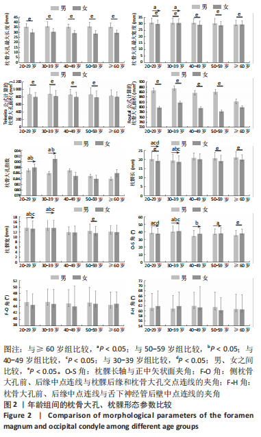

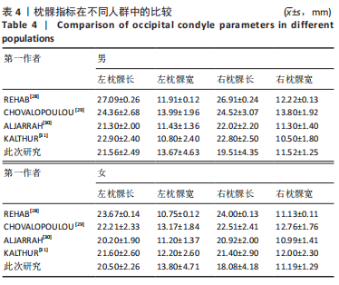
 3.2 关于枕髁解剖参数的分析 “枕-寰-枢关节”对寰枕关节的稳定性起重要作用。枕髁的大小和方向可能影响颅颈交界区病变的手术入路[25]。对于枕骨大孔腹侧和斜坡处的病变可经远外侧入路暴露,术中磨除枕髁一定范围后可扩大手术视野和增大操作空间,但结果是可能导致枕颈不稳,因此,就是否磨除枕髁及磨除范围的大小是目前学者争议较大的地方,但总体来讲要根据具体情况考虑是否磨除枕髁。WANIBUCHI等[26]认为在远外侧入路中枕骨大孔周围骨质磨除范围应在保持寰枕关节稳定的原则下采取个体化设计,此外置钉的成功与还否取决于枕髁体积的大小。此次研究中发现枕髁长和枕髁宽具有左右侧别和性别差异,在枕髁长中整体男性左侧大于女性,且右侧小于左侧;枕髁宽男性右侧大于女性,男性左侧小于女性,且左侧大于右侧。说明枕髁的长、宽外形上存在着不对称性;男性枕髁可容纳较大的螺钉,同时术中处理枕髁时需考虑性别、侧别带来的差异性[27]。REHAB等[28]、CHOVALOPOULOU等[29]、ALJARRAH等[30]、KALTHUR等[31]结果都与此次研究不同,见表4,可能与样本来自不同国家、地区及测量方法有关,需要对检测手段、颅底标志制定相关标准。
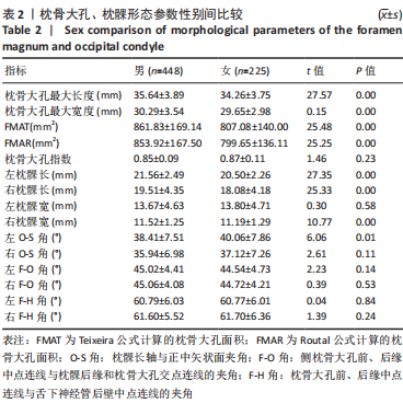
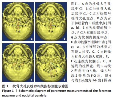
 此次研究中所测量的角度中,只有左O-S角有性别间的统计学差异,男性O-S角小于女性;只有F-H角无侧别间统计学差异,左侧O-S角大于右侧,左侧F-O角小于右侧。O-S角大小反映枕髁后缘与正中矢状面的距离,角度越大手术视野也越大,在磨除枕骨后左侧视野大于右侧。F-O角为髁后入路不需磨除枕髁时所暴露的手术视野范围,在磨除枕骨至枕髁后缘后所暴露的范围左侧小于右侧,建议从右侧入路。ZHOU等[32]对27例CT测量结果左侧O-S角小于右侧,结果与此文一致。GUMUSSOY等[33]、KIRNAZ等[34]研究左侧O-S角大于右侧,不同于此次研究结果,可能因其样本量较少所致。F-H角为远外侧经髁入路时,从枕髁后缘磨除至舌下神经管后壁时所暴露的最大手术视野范围,此文结果为男性F-H角小于女性,且左侧小于右侧。经髁入路时女性右侧枕髁磨除至舌下神经管后所暴露的手术视野范围相对较大,但磨除枕髁应根据具体情况在枕髁后缘和舌下神经管后壁之间的范围内进行。胡海建[27]的结果中左侧F-H角大于右侧,男性F-H角大于女性,与此文不一致,可能由于样本来源不同导致枕髁形态与此次研究有差异。对于枕髁磨除多少,有国内外学者认为磨除枕髁后内侧1/3至1/2即可使肿瘤暴露满意,磨除枕髁的1/2以下基本不影响寰枕关节的稳定性[35-36]。