[1] ELSOE R, JOHANSEN MB, LARSEN P. Tibial plateau fractures are associated with a long-lasting increased risk of total knee arthroplasty a matched cohort study of 7,950 tibial plateau fractures. Osteoarthritis Cartilage. 2019;27(5):805-809.
[2] GASTON P, WILL EM, KEATING JF. Recovery of knee function following fracture of the tibial plateau. J Bone Joint Surg Br. 2005;87-B(9):1233-1236.
[3] HOHL M. Tibial condylar fractures. J Bone Joint Surg Am. 1967;49-A(7): 1455-1467.
[4] MOORE TM. Fracture-dislocation of the knee. Clin Orthop Relat Res. 1981;156:128-140.
[5] SCHATZKER J. Compression in the surgical treatment of fractures of the tibia. Clin Orthop Relat Res. 1974;105:220-239.
[6] MARSH JL, SLONGO TF, AGEL J, et al. Fracture and dislocation classification compendium : orthopaedic Trauma Association classification, database andoutcomes committee. J Orthop Trauma. 2007;21(10 Suppl):1-133.
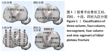
[7] LUO CF, SUN H, ZHANG B, et al. Three-column fixation for complex tibial plateau fractures. J Orthop Trauma. 2010;24(11):683-692.
[8] 张彬彬,罗从风,王驭恺,等.胫骨平台骨折手术治疗的新三柱理念[J].上海医学,2017,40(6):341-348.
[9] CHANG SM. Selection of surgical approaches to the posterolateral tibial plateau fracture by its combination patterns. J Orthop Trauma. 2011;25(3):32-33.
[10] KRAUSE M, PREISS A, MÜLLER G, et al. Intra-Articular Tibial Plateau Fracture Characteristics according to the “Ten Segment Classification”. Injury. 2016;47(11):2551-2557.
[11] YAO X, XU Y, YUAN J, et al. Classification of tibia plateau fracture according to the “four-column and nine-segment”. Injury. 2018;49(12): 2275-2283.
[12] MOLENAARS RJ, MELLEMA JJ, DOORNBERG JN, et al. Tibial Plateau Fracture Characteristics: Computed Tomography Mapping of Lateral, Medial and Bicondylar Fractures. J Bone Joint Surg Am. 2015;97(18): 1512-1520.
[13] YAO X, ZHOU KH, LV B, et al. 3D mapping and classification of tibial plateau fractures. Bone Joint Res. 2020;9(6):258-267.
[14] MEULENKAMP B, MARTIN R, DESY NM, et al. Incidence,Risk Factors, and Location of Articular Malreductions of the Tibial Plateau. J Orthop Trauma. 2017;31(3):146-150.
[15] HOEKSTRA H, KEMPENAERS K, NIJS S. A revised 3-column classification approach for the surgical planning of extended lateral tibial plateau fractures. Eur J Trauma Emerg Surg. 2017;43(5):637-643.
[16] KRAUSE M, PREISS A, MEENEN NM, et al. Fracturoscopy is superior to fluoroscopy in the articular reconstruction of complex tibial plateau fractures - an arthroscopy assisted fracture reduction technique. J Orthop Trauma. 2016;30(8):437-444.
[17] YANG G, ZHU Y, LUO C, et al. Morphological characteristics of Schatzker type IV tibial plateau fractures: a computer tomography based study. Int Orthop. 2012;36(11):2355-2360.
[18] ZHAI Q, HU C, XU Y, et al. Morphologic Study of Posterior Articular Depression in Schatzker IV Fractures. Orthopedics. 2015;38(2):124-128.
[19] COLE PA, MEHRLE RK, BHANDARI M, et al. The pilon map: fracture lines and comminution zones in OTA/AO type 43C3 pilon fractures. J Ortho Trauma. 2013;27(7):e152-156.
[20] 赵腾飞,汤欣.三维重建绘制胫骨平台骨折线地图及分析[D].大连:大连医科大学,2017.
[21] MCGONAGLE L, CORDIER T, LINK BC, et al. Tibia plateau fracture mapping and its influence on fracture fixation. J Orthop Trauma. 2019; 20(12):1-6.
[22] TSCHERNE H, LOBENHOFFER P. Tibial plateau fractures:Management and expected results. Clin Orthop. 1993;292:87-100.
[23] CHANG SM, ZHANG YQ, YAO MW, et al. Schatzker Type IV Medial Tibial Plateau Fractures: A Computed Tomography-based Morphological Subclassification. Orthopedics (Online). 2014;37(8):699-706.
[24] 马卓,张世民,胡孙君,等.Schatzker Ⅳ型胫骨平台双髁骨折的CT亚型分类及临床意义[J].中华创伤骨科杂志,2016,18(10):832-839.
[25] KFURI M, SCHATZKER J. Revisiting the Schatzker classification of tibial plateau fractures. Injury. 2018;49(12):2252-2263.
[26] WAHLQUIST M, IAGUILLI N, EBRAHEIM N, et al. Medial Tibial Plateau Fractures: a New Classification System. J Trauma. 2007;63(6):1418-1421.
[27] PURNELL ML, LARSON AI, SCHNETZLER KA, et al. Diagnosis and Surgical Treatment of Schatzker Type IV Variant Biplanar Medial Tibial Plateau Fractures in Alpine Skiers. Tech Knee Surg. 2007;6(1):17-28.
[28] 于吉文,刘建,何维栋,等.根据CT扫描及三维重建改良胫骨平台骨折的Schatzker分型[J].实用骨科杂志,2011,17(1): 28-32.
[29] 杨光,田书建,高延征,等.CT评价Schatzker Ⅳ型胫骨平台骨折的形态学特点[J].中华实用诊断与治疗杂志,2016,30(9):891-893.
[30] 罗从风,姜锐,周曼瑜,等.胫骨内侧平台骨折手术治疗失败的原因分析[J].中华创伤骨科杂志,2006,8(7):642-646.
[31] KAPLAN PA, GEHL RH, DUSSAULT RG, et al. Bone contusions of the posterior lip of the medial tibial plateau (contrecoup injury) and associated internal derangements of the knee at MR imaging. Radiology. 1999;211(3):747-753.
[32] WYMENGA AB, KATS JJ, KOOLOOS J, et al. Surgical anatomy of the medial collateral ligament and the posteromedial capsule of the knee. Knee Surg Sports Traumatol Arthrosc. 2006;14(3):229-234.
[33] SATTERWHITE Y. The anatomy and biomechanics of the medial structures of the knee. Oper Tech Sports Med. 1996;4(3):134-140.
[34] ROBINSON JR, SANCHEZ-BALLESTER J, BULL AM, et al. The posteromedial corner revisited. J Bone Joint Surg Br. 2004;86(5):674-681.
|
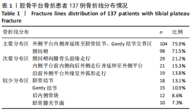
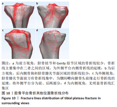
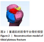
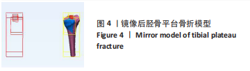
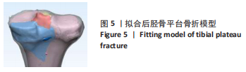
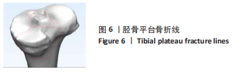
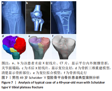
 研究回顾性分析137例胫骨平台骨折患者的病历资料,以原始骨折CT数据为基础,重建并虚拟复位平台骨折三维模型,描述骨折线特点,揭示其走行与分布规律,以期指导临床治疗。