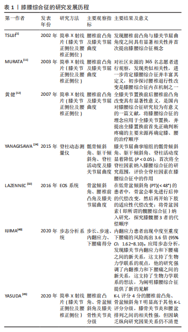
[1] TSUJI T, MATSUYAMA Y, GOTO M, et al. Knee-spine syndrome: correlation between sacral inclination and patellofemoral joint pain. J Orthop Sci. 2002; 7(5):519-523.
[2] UEHARA K, AKAI M, DOI T, et al. Relationship between X-ray findings of lumbar spondylosis and knee pain. BMC Musculoskeletal Disorders. 2019; 20(1):379.
[3] BLAGOJEVIC M, JINKS C, JEFFERY A, et al. Risk factors for onset of osteoarthritis of the knee in older adults: a systematic review and meta-analysis. Osteoarthritis Cartilage. 2010;18(1):24-33.
[4] BURGER H, VAN DAELE PL, ODDING E, et al. Association of radiographically evident osteoarthritis with higher bone mineral density and increased bone loss with age. The Rotterdam Study. Arthritis Rheum. 1996;39(1):81-86.
[5] DAVIS MA, ETTINGER WH, NEUHAUS JM, et al. The association of knee injury and obesity with unilateral and bilateral osteoarthritis of the knee. Am J Epidemiol. 1989;130(2):278-288.
[6] HART DJ, DOYLE DV, SPECTOR TD. Incidence and risk factors for radiographic knee osteoarthritis in middle-aged women: the Chingford Study. Arthritis Rheum. 1999;42(1):17-24.
[7] MURAKI S, AKUNE T, OKA H, et al. Incidence and risk factors for radiographic knee osteoarthritis and knee pain in Japanese men and women: a longitudinal population-based cohort study. Arthritis Rheum. 2012;64(5):1447-1456.
[8] SEKI T, HASEGAWA Y, YAMAGUCHI J, et al. Association of serum carotenoids, retinol, and tocopherols with radiographic knee osteoarthritis: possible risk factors in rural Japanese inhabitants. J Orthop Sci. 2010;15(4):477-484.
[9] YUDOH K, NGUYEN V, NAKAMURA H, et al. Potential involvement of oxidative stress in cartilage senescence and development of osteoarthritis: oxidative stress induces chondrocyte telomere instability and downregulation of chondrocyte function. Arthritis Res Ther. 2005;7(2):R380-R391.
[10] OFFIERSKI CM, MACNAB I. Hip-spine syndrome. Spine (Phila Pa 1976). 1983; 8(3):316-321.
[11] MURATA Y, TAKAHASHI K, YAMAGATA M, et al. The knee-spine syndrome. The Journal of Bone and Joint Surgery. British Volume. 2003;85-B(1):95-99.
[12] 黄健,吕厚山,白楚杰,等.膝关节骨性关节炎合并退行性变性腰椎病变全膝关节置换术后患者腰椎前凸角度与膝关节屈曲矫正度的变化[J].中国组织工程研究与临床康复,2007,11(4):722-724.
[13] 郑文鑫.腰椎管狭窄手术前后CT测量椎管容积的价值研究[D].海口:海南医学院,2018.
[14] 戴力杨.腰椎屈伸活动对椎管容量的影响[J].第二军医大学学报,1989, 9(3):197-199.
[15] GLASSMAN SD, BRIDWELL K, DIMAR JR, et al. The impact of positive sagittal balance in adult spinal deformity. Spine (Phila Pa 1976). 2005;30(18): 2024-2029.
[16] MIN K, HAHN F, LEONARDI M. Lumbar spinal osteotomy for kyphosis in ankylosing spondylitis: the significance of the whole body kyphosis angle. J Spinal Disord Tech. 2007;20(2):149-153.
[17] KECHAGIAS VA, GRIVAS TB, PAPAGELOPOULOS PJ, et al. Investigation of the relationship between hip and knee osteoarthritis and disordered spinal and pelvic morphology. Cureus. 2022;14(1):e20861.
[18] WANG WJ, LIU F, ZHU YW, et al. Sagittal alignment of the spine-pelvis-lower extremity axis in patients with severe knee osteoarthritis. Bone Joint Res. 2016,5(5):198-205.
[19] 刘飞,王渭君,翁文杰,等.膝关节骨关节炎患者脊柱-骨盆-下肢矢状面形态变化的初步研究[J].中国矫形外科杂志,2015,23(9):784-789.
[20] LEE CS, PARK SJ, CHUNG SS, et al. The effect of simulated knee flexion on sagittal spinal alignment: novel interpretation of spinopelvic alignment. Eur Spine J. 2013;22(5):1059-1065.
[21] KATSUMI R, MANNEN EM, BAJAJ G, et al. The influence of knee osteoarthritis on spinopelvic alignment and global sagittal balance. J Knee Surg. 2022. doi: 10.1055/s-0042-1747947.
[22] 张胜国,刘海鹰,王波,等.膝关节屈曲畸形与腰椎和骨盆矢状位对线的相关性研究[J].中国矫形外科杂志,2013,21(9):914-917.
[23] TAUCHI R, IMAGAMA S, MURAMOTO A, et al. Influence of spinal imbalance on knee osteoarthritis in community-living elderly adults. Nagoya J Med Sci. 2015;77(3):329-337.
[24] YANAGISAWA S, SATO N, SHIMIZU M, et al. Relation among the knee, sagittal spinal alignment, and the spinal range of motion: investigation in local medical check-ups using the Spinal Mouse. Asia Pac J Sports Med Arthrosc Rehabil Technol. 2015;2(2):68-71.
[25] HARATO K, NAGURA T, MATSUMOTO H, et al. A gait analysis of simulated knee flexion contracture to elucidate knee-spine syndrome. Gait Posture. 2008;28(4):687-692.
[26] IMAGAMA S, ITO Z, WAKAO N, et al. Influence of spinal sagittal alignment, body balance, muscle strength, and physical ability on falling of middle-aged and elderly males. Eur Spine J. 2013;22(6):1346-1353.
[27] IMAGAMA S, MATSUYAMA Y, HASEGAWA Y, et al. Back muscle strength and spinal mobility are predictors of quality of life in middle-aged and elderly males. Eur Spine J. 2011;20(6):954-961.
[28] POST RB, LEFERINK VJ. Spinal mobility: sagittal range of motion measured with the Spinal Mouse, a new non-invasive device. Arch Orthop Trauma Surg. 2004;124(3):187-192.
[29] FUNAO H, TSUJI T, HOSOGANE N, et al. Comparative study of spinopelvic sagittal alignment between patients with and without degenerative spondylolisthesis. Eur Spine J. 2012;21(11):2181-2187.
[30] BARREY C, ROUSSOULY P, LE HUEC JC, et al. Compensatory mechanisms contributing to keep the sagittal balance of the spine. Eur Spine J. 2013;22 Suppl 6:S834-S841.
[31] 李青,翁文杰,王渭君,等.EOS成像系统的介绍及其评估下肢力线临床价值的研究现状[J].中国骨伤,2019,32(9):875-878.
[32] LAZENNEC J Y, FOLINAIS D, BENDAYA S, et al. The global alignment in patients with lumbar spinal stenosis: our experience using the EOS full-body images. Eur J Orthop Surg Traumatol. 2016;26(7):713-724.
[33] OBEID I, HAUGER O, AUNOBLE S, et al. Global analysis of sagittal spinal alignment in major deformities: correlation between lack of lumbar lordosis and flexion of the knee. European Spine Journal. 2011;20(S5):681-685.
[34] FERRERO E, LIABAUD B, CHALLIER V, et al. Role of pelvic translation and lower-extremity compensation to maintain gravity line position in spinal deformity. J Neurosurg Spine. 2016;24(3):436-446.
[35] 冯其金,赵玲娟,郑昆仑,等.有限元分析法在腰椎生物力学中的研究进展[J].中国中西医结合外科杂志,2018,24(2):255-258.
[36] 吕震.膝关节退变对KLS腰椎运动单元负荷改变机制的研究[D].天津:天津医科大学,2019.
[37] ADAMS MA, HUTTON WC. The effect of posture on the role of the apophysial joints in resisting intervertebral compressive forces. J Bone Joint Surg Br. 1980;62(3):358-362.
[38] 周恩昌,唐萍,殷浩,等.膝内翻对骨盆─腰椎矢状位序列影响的有限元分析[J].中国骨与关节损伤杂志,2017,32(12):1233-1236.
[39] 谈绎文,郑昱新,詹红生,等.三维步态分析在膝骨关节炎研究中的应用[J].国际骨科学杂志,2014,35(4):215-218.
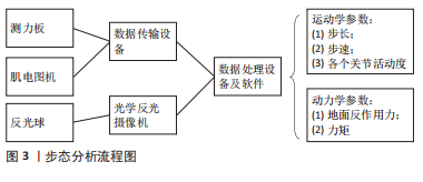
[40] IIJIMA H, SUZUKI Y, AOYAMA T, et al. Relationship between varus thrust during gait and low back pain in individuals with knee osteoarthritis. Arthritis Care Res. 2020;72(9):1231-1238.
[41] CHANG A, HAYES K, DUNLOP D, et al. Thrust during ambulation and the progression of knee osteoarthritis. Arthritis Rheum. 2004;50(12):3897-3903.
[42] FUKUTANI N, IIJIMA H, FUKUMOTO T, et al. Association of varus thrust with pain and stiffness and activities of daily living in patients with medial knee osteoarthritis. Phys Ther. 2016;96(2):167-175.
[43] IIJIMA H, FUKUTANI N, AOYAMA T, et al. Clinical phenotype classifications based on static varus alignment and varus thrust in japanese patients with medial knee osteoarthritis. Arthritis Rheumatol. 2015;67(9):2354-2362.
[44] SOTELO M, EICHELBERGER P, FURRER M, et al. Walking with an induced unilateral knee extension restriction affects lower but not upper body biomechanics in healthy adults. Gait Posture. 2018;65:182-189.
[45] YASUDA T, TOGAWA D, HASEGAWA T, et al. Relationship between knee osteoarthritis and spinopelvic sagittal alignment in volunteers over 50 years of age. Asian Spine J. 2020;14(4):495-501.
[46] OSHIMA Y, WATANABE N, IIZAWA N, et al. Knee–hip–spine syndrome: improvement in preoperative abnormal posture fo llowing total knee arthroplasty. Adv Orthop. 2019;2019:1-9.
|