中国组织工程研究 ›› 2023, Vol. 27 ›› Issue (15): 2339-2343.doi: 10.12307/2023.621

• 干细胞基础实验 basic experiments of stem cells • 上一篇    下一篇

SD乳鼠原代皮质神经元及星形胶质细胞同时培养的实验方法

明  江1,廖益东1,宋文学1,穆德勇1,刘宗伟1,徐卡娅2   

  1. 1贵州医科大学,贵州省贵安新区   550025;2贵州医科大学附属医院神经外科,贵州省贵阳市   550004
  • 收稿日期:2022-06-22 接受日期:2022-07-25 出版日期:2023-05-28 发布日期:2022-10-17
  • 通讯作者: 徐卡娅,博士,主任医师,贵州医科大学附属医院神经外科,贵州省贵阳市 550004
  • 作者简介:明江,男,1996年生,贵州省从江县人,侗族,贵州医科大学在读硕士,主要从事间充质干细胞移植治疗缺血性脑卒中的研究。
  • 基金资助:
    国家自然科学基金项目(81901173,82060231),项目负责人:徐卡娅;贵州省普通高等学校青年科技人才成长项目(黔教合 KY字[2021]190),项目负责人:徐卡娅;贵州医科大学2018年度学术新苗培养及创新探索专项项目(黔科合平台人才[2018]5779-44),项目负责人:徐卡娅

Experimental method of simultaneous culture of primary cortical neurons and astrocytes of Sprague-Dawley suckling rats

Ming Jiang1, Liao Yidong1, Song Wenxue1, Mu Deyong1, Liu Zongwei1, Xu Kaya2   

  1. 1Guizhou Medical University, Guiyang 550025, Guizhou Province, China; 2Department of Neurosurgery, Affiliated Hospital of Guizhou Medical University, Guiyang 550004, Guizhou Province, China
  • Received:2022-06-22 Accepted:2022-07-25 Online:2023-05-28 Published:2022-10-17
  • Contact: Xu Kaya, MD, Chief physician, Department of Neurosurgery, Affiliated Hospital of Guizhou Medical University, Guiyang 550004, Guizhou Province, China
  • About author:Ming Jiang, Master candidate, Guizhou Medical University, Guiyang 550025, Guizhou Province, China
  • Supported by:
    the National Natural Science Foundation of China, No. 81901173, 82060231 (to XKY); General Colleges and Universities Youth Science and Technology Talent Growth Project of Guizhou Province, No. [2021]190 (to XKY); 2018 Special Project for Academic Seedling Cultivation and Innovation Exploration of Guizhou Medical University, No. [2018]5779-44 (to XKY)

摘要:

文题释义:
原代皮质神经元:是神经系统的重要结构和功能单位,在神经生物学中扮演着重要的角色。此类细胞取材容易,可直接从新鲜SD乳鼠脑组织中提取,而且其特性接近于体内神经元特性,可作为细胞模型用于研究神经系统相关疾病,如缺血性脑卒中、糖尿病性脑病、神经退行性变化等。
星形胶质细胞:作为神经系统中最丰富的细胞类型,执行着重要的稳态功能。病理状态下星形胶质细胞的活化,几乎参与所有形式的神经损伤和神经性疾病。因此获取稳定的星形胶质细胞模型,对于研究星形胶质细胞引发的相关疾病具有重要意义。

背景:目前获取原代皮质神经元和星形胶质细胞的方法很多,传统方法通常是分别获取这两种细胞,但实验方法过于繁琐且浪费实验材料。找到一种简单、经济、可行并同时提取这两种细胞的培养方法尤为重要。
目的:观察同时提取培养SD乳鼠大脑皮质神经元及星形胶质细胞的效果及注意事项。
方法:选用出生24 h内的SD乳鼠,用体积分数为75%乙醇浸泡消毒,待乳鼠昏迷后断脊处死,沿乳鼠颈部用剪刀离断头颅并放入装有高糖DMEM的小烧杯中,沿矢状缝打开头颅并取出大脑放入盛有高糖DMEM的培养皿中。在冰上剥出脑膜及血管,夹取皮质表面2.0-3.0 mm的脑组织,通过木瓜酶和DNA酶进行消化20 min,用含有体积分数为10%胎牛血清的DMEM/F12终止消化。以细胞浓度为1.0×109 L-1种植于含有爬片(用多聚赖氨酸处理)的6孔板中,分别于1,3,5,7 d通过倒置荧光显微镜下观察神经元的形态变化,使用Calcien-AM进行活细胞染色观察细胞活性,β-Tubulin、MAP2免疫荧光染色对神经元进行鉴定。将剩余大脑皮质进行星形胶质细胞的提取,经过胰酶消化、过滤、吹打,以适宜的浓度接种于培养瓶中,待细胞融合度达到90%时,进行恒温水平摇床振荡24 h以纯化星形胶质细胞。当细胞传代纯化后以1.0×10⁹ L-1接种于6孔板中培养,GFAP免疫荧光染色对星形胶质细胞进行鉴定。
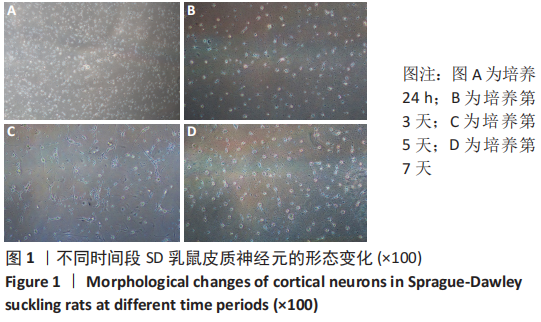
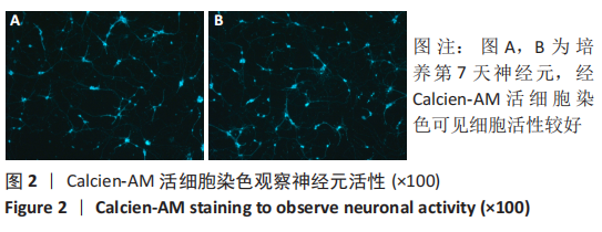
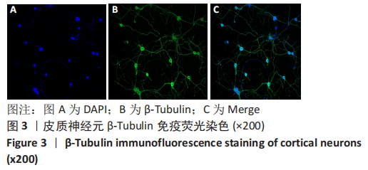
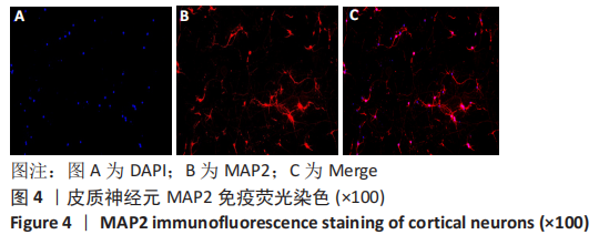
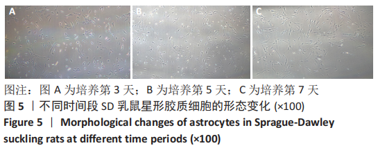
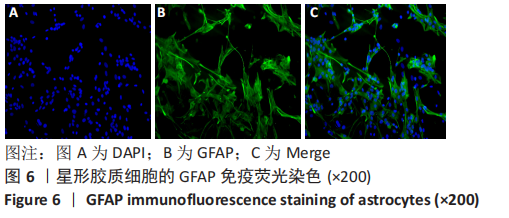
结果与结论:①培养1 d后可见神经元贴壁生长,胞体增大,部分聚集生长,细胞间有少量的突触连接;培养3,5 d后胞体进一步增大,突触增粗伸长相互连接;培养7 d后神经元明显聚集,突触增长增粗,相互连接形成致密的细胞网。Calcien-AM活细胞染色可见神经元活性较好,经β-Tubulin、MAP2免疫荧光鉴定细胞纯度大于90%。②培养3 d后星形胶质细胞胞体增大,呈现梭状、不规则状,细胞间出现少量相互连接,细胞间存在较多小胶质细胞;培养5 d后胞体、突起进一步增大;培养7 d发现细胞间的小胶质细胞及其他杂质明显减少,背景较为干净。经GFAP免疫荧光鉴定细胞纯度大于95%。③此方法获得的皮质神经元和星形胶质细胞形态好、杂质少、纯度高,可满足神经科学研究对高纯度的皮质神经元及星形胶质细胞的需求。
https://orcid.org/0000-0002-7244-663X (明江) 

中国组织工程研究杂志出版内容重点:干细胞;骨髓干细胞;造血干细胞;脂肪干细胞;肿瘤干细胞;胚胎干细胞;脐带脐血干细胞;干细胞诱导;干细胞分化;组织工程

关键词: 皮质神经元, 星形胶质细胞, DNA酶, 免疫荧光, 原代培养

Abstract: BACKGROUND: At present, there are many methods to obtain primary cortical neurons and astrocytes. The traditional method is usually to obtain these two kinds of cells separately, but the experimental method is too cumbersome and wastes experimental materials. It is particularly important to find a simple, economical, and feasible culture method that simultaneously extracts both types of cells.  
OBJECTIVE: To observe the effect and precautions of simultaneously extracting neurons and astrocytes from the cerebral cortex of Sprague-Dawley suckling rats.
METHODS: The suckling rats within 24 hours of birth were selected and sterilized with 75% alcohol. After the suckling rats were in a coma, they were sacrificed by cutting their spines. The head was severed with scissors along the neck of the suckling rats and placed in a small beaker containing high-glucose DMEM. The skull was opened through the suture and the brain was removed and placed in a petri dish containing high glucose DMEM. The meninges and blood vessels were peeled off on ice, and the brain tissue 2.0-3.0 mm on the surface of the cortex was clipped, digested with papain and DNase, and terminated with DMED/F12 containing 10% fetal bovine serum. Samples were seeded at a density of 1.0×10⁹/L in six-well plates containing slides (treated with polylysine). The morphological changes of neurons were observed under an inverted fluorescence microscope at 1, 3, 5, and 7 days, respectively. Calcien-AM was used for live cell staining to observe cell viability, and β-Tubulin and MAP2 immunofluorescence staining was used to identify neurons. Astrocytes were extracted from the remaining cerebral cortex. After trypsin digestion, filtration, and pipetting, they were inoculated into culture flasks at a suitable concentration. When the cell confluency reached 90%, the astrocytes were shaken on a constant temperature horizontal shaker for 24 hours to purify the astrocytes. After the cells were passaged and purified, they were seeded in six-well plates at 1.0×10⁹/L. Astrocytes were identified by GFAP immunofluorescence staining. 
RESULTS AND CONCLUSION: (1) After 1 day of culture, neurons could be seen to adhere to the wall; the cell body increased, and some of the cells grew together, and there were a few synaptic connections between cells. After 3 and 5 days, the cell body further increased, and the synapses were thickened and elongated to form a sparse network. After 7 days, the neurons were obviously aggregated; the synapses grew and thickened, and they connected to each other to form a dense cell network. Calcien-AM live cell staining showed better neuronal viability. The purity of the two identification methods was greater than 90% identified by β-Tubulin and MAP2 immunofluorescence staining. (2) After 3 days of culture, the astrocyte bodies were enlarged, showing spindle and irregular shapes, with a small amount of interconnected cells and many microglia between cells. After 5 days of culture, the cell bodies and protrusions further increased. After 7 days of culture, it was found that the microglia and other impurities between the cells were reduced compared with the previous ones, and the background was relatively clean. The purity of the cells was greater than 95% identified by GFAP immunofluorescence staining. (3) The obtained cortical neurons and astrocytes have good morphology, less impurities and high purity. They can meet the needs of high-purity cortical neurons and astrocytes for neuroscience research.

Key words: cortical neuron, astrocyte, DNase, immunofluorescence, primary culture

中图分类号: