[1] NAULT ML, ALLARD P, HINSE S, et al. Relations between standing stability and body posture parameters in adolescent idiopathic scoliosis. Spine (Phila Pa 1976). 2002;27(17):1911-1917.
[2] EL HAWARY R, CHUKWUNYERENWA C. Update on evaluation and treatment of scoliosis. Pediatr Clin North Am. 2014;61(6):1223-1241.
[3] 陈绍丰,李博,周潇逸,等.青少年特发性脊柱侧凸相关生活质量评价量表现状[J].第二军医大学学报,2020,41(11):1188-1192.
[4] WEISS HR, MORAMARCO M. Indication for surgical treatment in patients with adolescent Idiopathic Scoliosis-a critical appraisal. Patient Saf Surg. 2013;7(1):17.
[5] LUAN FJ, WAN Y, MAK KC, et al. Cancer and mortality risks of patients with scoliosis from radiation exposure: a systematic review and meta-analysis. Eur Spine J. 2020;29(12):3123-3134.
[6] LEVY AR, GOLDBERG MS, MAYO NE, et al. Reducing the lifetime risk of cancer from spinal radiographs among people with adolescent idiopathic scoliosis. Spine (Phila Pa 1976). 1996;21(13):1540-1547.
[7] RONCKERS CM, LAND CE, MILLER JS, et al. Cancer mortality among women frequently exposed to radiographic examinations for spinal disorders. Radiat Res. 2010;174(1):83-90.
[8] RONCKERS CM, DOODY MM, LONSTEIN JE. Multiple diagnostic X-rays for spine deformities and risk of breast cancer. Cancer Epidemiol Biomarkers Prev. 2008;17(3):605-613.
[9] HOFFMAN DA, LONSTEIN JE, MORIN MM, et al. Breast cancer in women with scoliosis exposed to multiple diagnostic X rays. J Natl Cancer Inst. 1989;81(17):1307-1312.
[10] OAKLEY PA, EHSANI NN, HARRISON DE. The scoliosis quandary: are radiation exposures from repeated X-rays harmful? Dose Response. 2019;17(2):1559325819852810.
[11] DESCHÊNES S, CHARRON G, BEAUDOIN G, et al. Diagnostic imaging of spinal deformities: reducing patients radiation dose with a new slot-scanning X-ray imager. Spine (Phila Pa 1976). 2010;35(9):989-994.
[12] BRANCHINI M, DEL VECCHIO A, GIGLIOTTI CR. Organ doses and lifetime attributable risk evaluations for scoliosis examinations of adolescent patients with the EOS imaging system. Radiol Med. 2018;123(4):305-313.
[13] SUZUKI S, YAMAMOTO T, SHIKATA J, et al. Ultrasound measurement of vertebral rotation in idiopathic scoliosis. J Bone Joint Surg Br. 1989; 71:252-255.
[14] KWOK G, YIP J, YICK KL, et al. postural screening for adolescent idiopathic scoliosis with infrared thermography. Sci Rep. 2017;7(1):14431.
[15] VASILIADIS E, GRIVAS TB. Quality of life after conservative treatment of adolescent idiopathic scoliosis. Stud Health Technol Inform. 2008; 135:409-413.
[16] 中华人民共和国国家卫生和计划生育委员会、中国国家标准化管理委员会.儿童青少年脊柱弯曲异常的筛查:GB/T 16133-2014[S]. 2014.
[17] 中国青少年脊柱侧凸筛查临床实践指南及路径指引[J].中华骨科杂志,2020,40(23):1574-1582.
[18] CÔTÉ P, KREITZ BG, CASSIDY JD, et al. A study of the diagnostic accuracy and reliability of the Scoliometer and Adam’s forward bend test. Spine (Phila Pa 1976). 1998;23(7):796-803.
[19] VIVIANI GR, BUDGELL L, DOK C, et al. Assessment of accuracy of the scoliosis school screening examination. Am J Public Health. 1984;74(5): 497-498.
[20] SENKOYLU A, ILHAN MN, ALTUN N, et al. A simple method for assessing rotational flexibility in adolescent idiopathic scoliosis: modified Adam’s forward bending test. Spine Deform. 2021;9(2):333-339.
[21] FRANKO OI, BRAY C, NEWTON PO. Validation of a scoliometer smartphone app to assess scoliosis. J Pediatr Orthop. 2012;32(8):72-75.
[22] Negrini S, Donzelli S, Di Felice F, et al. Construct validity of the trunk aesthetic clinical evaluation (TRACE) in young people with idiopathic scoliosis. Ann Phys Rehabil Med. 2020;63(3):216-221.
[23] NAVARRO IJ, CANDOTTI CT, FURLANETTO TS, et al. Validation of a mathematical procedure for the cobb angle assessment based on photogrammetry. J Chiropr Med. 2019;18(4):270-277.
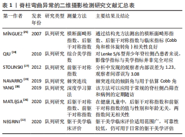
[24] QIU Y, QIU XS, MA WW, et al. How well do radiological measurements correlate with cosmetic indices in adolescent idiopathic scoliosis with Lenke 5, 6 curve types? Spine. 2010;35(18):882-888.
[25] MÍNGUEZ MF, BUENDÍA M, CIBRIÁN RM, et al. Quantifier variables of the back surface deformity obtained with a noninvasive structured light method: evaluation of their usefulness in idiopathic scoliosis diagnosis. Eur Spine J. 2007;16(1):73-82.
[26] MATLĘGA A, STĘPOWSKA J, WIŚNIEWSKI A, et al. Assessment of the coronal plane trunk symmetry in children. Phys Theory Pract. 2020; 36(12):1502-1508.
[27] STOLINSKI L, KOTWICKI T, CZAPROWSKI D, et al. Analysis of the Anterior Trunk Symmetry Index (ATSI). Preliminary report. Stud Health Technol Inform. 2012;176:242-246.
[28] ZHANG Y, SHI J, PENG Y, et al. Artificial intelligence-enabled screening for diabetic retinopathy: a real-world, multicenter and prospective study. BMJ Open Diabetes Res Care. 2020;8(1):e001596.
[29] GUROVICH Y, HANANI Y, BAR OY, et al. Identifying facial phenotypes of genetic disorders using deep learning. Nat Med. 2019;25(1):60-64.
[30] YANG J, ZHANG K, FAN H, et al. Development and validation of deep learning algorithms for scoliosis screening using back images. Commun Biol. 2019;25(2):390.
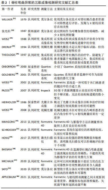
[31] WILLNER S. Moiré topography for the diagnosis and documentation of scoliosis. Acta Orthop Scand. 1979;50(3):295-302.
[32] SATO T, YONEZAWA I, AKIMOTO T, et al. Novel hump measurement system with a 3D camera for early diagnosis of patients with adolescent idiopathic scoliosis: a study of accuracy and reliability. Cureus. 2020; 12(5):e8229.
[33] 张光铂,李子荣,魏新荣,等.波纹照相在脊柱侧凸普查中的应用[J].中华外科杂志,1987,25(7):387-389.
[34] HIERHOLZER E, SCHIER F. Rasterstereography in the measurement and postoperative follow-up of anterior chest wall deformities. Z Kinderchir. 1986;41(5):267-271.
[35] WEISZ I, JEFFERSON RJ, TURNER-SMITH AR, et al. ISIS scanning: a useful assessment technique in the management of scoliosis. Spine. 1988; 13:405-408.
[36] THEOLOGIS T, FAIRBANK JCT, TURNER-SMITH A, et al. Early detection of progression in adolescent idiopathic scoliosis by measurement of changes in back shape with the integrated shape imaging system scanner. Spine. 1997;22(11):1223-1227.
[37] OXBORROW N. Assessing the child with scoliosis: the role of surface topography. Arch Dis Child. 2000;83:453-455.
[38] GOLDBERG CJ, KALISZER M, MOORE DP, et al. Surface topography, Cobb angles, and cosmetic change in scoliosis. Spine (Phila Pa 1976). 2001;26(4):E55-E63.
[39] DRERUP B, HIERHOLZER E. Back shape measurement using video rasterstereography and three-dimensional reconstruction of spinal shape. Clin Biomech. 1994;9(1):28-36.
[40] WEISS HR, ELOBEIDI N. Comparison of the kyphosis angle evaluated by video rasterstereography (VRS) with x-ray measurements. Stud Health Technol Inform. 2008;140:137-139.
[41] WEISS HR, DIECKMANN J, GERNER HJ. Outcome of in-patient rehabilitation in patients with M. Scheuermann evaluated by surface topography. Stud Health Technol Inform. 2002;88:246-249.
[42] SCHROEDER J, REER R, BRAUMANN KM. Video raster stereography back shape reconstruction: a reliability study for sagittal, frontal, and transversal plane parameters. Eur Spine J. 2015;24(2):262-269.
[43] PAZOS V, CHERIET F, DANSERAU J, et al. Reliability of trunk shape measurements based on 3-D surface reconstructions. Eur Spine J. 2007;16(11):1882-1891.
[44] HONG A, JASWAL N, WESTOVER L, et al. surface topography classification trees for assessing severity and monitoring progression in adolescent idiopathic scoliosis. Spine (Phila Pa 1976). 2017;42(13): E781-E787.
[45] MANGONE M, RAIMONDI P, PAOLONI M, et al. Vertebral rotation in adolescent idiopathic scoliosis calculated by radiograph and back surface analysis-based methods: correlation between the Raimondi method and rasterstereography. Eur Spine J. 2013;22(2):367-371.
[46] APPLEBAUM A, CHO W, NESSIM A, et al. Establishing the validity of surface topography for assessment of scoliosis: a prospective study. Spine Deform. 2021;9(3):685-689.
[47] MICHALIK R, KNOD M, SIEBERS H, et al. Introduction and evaluation of a novel multi-camera surface topography system.Gait Posture. 2020; 80:367-373.
[48] BASSANI T, STUCOVITZ E, GALBUSERA F, et al. Is rasterstereography a valid noninvasive method for the screening of juvenile and adolescent idiopathic scoliosis? Eur Spine J. 2019;28(3):526-535.
[49] JAREMKO JL, PONCET P, RONSKY J, et al. Indices of torso asymmetry related to spinal deformity in scoliosis. Clin Biomech. 2002;17:559-568.
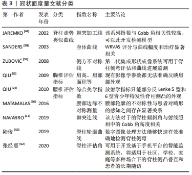
[50] SANDERS JO, POLLY DW JR, CATS-BARIL W, et al. Analysis of patient and parent assessment of deformity in idiopathic scoliosis using the Walter Reed Visual Assessment Scale. Spine (Phila Pa 1976). 2003;28(18): 2158-2163.
[51] ZUBOVIĆ A, DAVIES N, BERRYMAN F, et al. New method of Scoliosis Deformity Assessment: ISIS2 System. Stud Health Technol Inform. 2008; 140:157-160.
[52] 张经惠,沈林勇,宋薇,等.用于日常体表监测的三维脊柱形态测量技术[J].生物医学工程学杂志,2020,37(5):809-817.
[53] 陆俊,邢丽冬,钱志余,等.基于数字图像处理的脊柱侧弯快速检测系统[J].中国医疗器械杂志,2019,43(4):259-262.
[54] KOTWICKI T, NEGRINI S, GRIVAS TH, et al. Methodology of evaluation of morphology of the spine and the trunk in idiopathic scoliosis and other spinal deformities -6th SOSORT consensus paper. Scoliosis. 2009;4:26.
[55] QIU XS, MA WW, LI WG, et al. Discrepancy between radiographic shoulder balance and cosmetic shoulder balance in adolescent idiopathic scoliosis patients with double thoracic curve. Eur Spine J. 2009;18(1):45-51.
[56] MATAMALAS A, BAGÓ J, AGATA E, et al. Validity and reliability of photographic measures to evaluate waistline asymmetry in idiopathic scoliosis. Eur Spine J. 2016;25(10):3170-3179.
[57] GARDNER A, BERRYMAN F, PYNSENT P. What is the variability in shoulder, axillae and waist position in a group of adolescents? J Anat. 2017;231(2):221-228.
[58] BERRYMAN F, PYNSENT P, FAIRBANK J, et al. A new system for measuring 3D back shape in scoliosis. Eur Spine J. 2008;17:663-672.
[59] ASHER M, LAI SM, BURTON D, et al. The influence of spine and trunk deformity on preoperative idiopathic scoliosis patients’ health-related quality of life questionnaire responses. Spine (Phila Pa 1976). 2004; 29(8):861-868.
[60] PARENT EC, DAMARAJU S, HILL D, et al. Identifying the best surface topography parameters for detecting idiopathic scoliosis curve progression. Stud Health Technol Inform. 2010;158:78-82.
[61] MINGUEZ MF, BUENDÍA M, CIBRIÁN RM, et al. Quantifier variables of the back surface deformity obtained with a noninvasive structured light method: evaluation of their usefulness in idiopathic scoliosis diagnosis. Eur Spine J. 2007;16(1):73-82.
[62] FAIRBANK J. Historical perspective: william adams, the forward bending test, and the spine of gideon algernon mantell. Spine (Phila Pa 1976). 2004;29(17):1953-1955.
[63] BUNNELL WP. An objective criterion for scoliosis screening. J Bone Joint Surg Am. 1984;66(9):1381-1387.
[64] SUZUKI N, INAMI K, ONO T, et al. In: Research into Spinal Deformities 2. Stokes IAF, editor. Amsterdam: IOS Press; 1999. Analysis of posterior trunk symmetry index (POTSI) in Scoliosis: Part 1: 81-84.
[65] ZAINA F, NEGRINI S, ATANASIO S. TRACE (Trunk Aesthetic Clinical Evaluation), a routine clinical tool to evaluate aesthetics in scoliosis patients: development from the Aesthetic Index (AI) and repeatability. Scoliosis. 2009;4:3.
[66] 徐锡明,魏显招,等.青少年特发性脊柱侧凸学校筛查的进展及意义[J].脊柱外科杂志,2013,11(6):360-364.
[67] DRERUP B, HIERHOLZER E. Evaluation of frontal radiographs of scoliotic spines--Part I. Measurement of position and orientation of vertebrae and assessment of clinical shape parameters. J Biomech. 1992;25(11):1357-1362.
[68] DRERUP B, HIERHOLZER E. Evaluation of frontal radiographs of scoliotic spines--Part II. Relations between lateral deviation, lateral tilt and axial rotation of vertebrae. J Biomech. 1992;25(12):1443-1450.
[69] DRERUP B, HIERHOLZER E. Assessment of scoliotic deformity from back shape asymmetry using an improved mathematical model. Clin Biomech (Bristol, Avon). 1996;11(7):376-383.
[70] 牛升波,杨桓,杨明园,等.脊柱畸形的影像学参数测量及其研究进展[J].第二军医大学学报,2020,41(11):1177-1182.
[71] 蒋志成,徐慧琼,万宇辉,等.儿童青少年脊柱弯曲异常筛查研究进展[J].中国学校卫生,2021,42(2):312-315, 320.
[72] 吕东辉,顼超静,孙九爱.图像处理技术在脊柱侧弯检查中的应用[J].生物医学工程学杂志,2012,29(4):663-668.
|