[1] SINGH K, SHAH H, JOSEPH B, et al. Evolution of Legg-Calvé-Perthes disease following proximal femoral varus osteotomy performed in the avascular necrosis stage:a prospective study. J Child Orthop. 2020; 14(1):58-67.
[2] LEROUX J, ABU AMARA S, LECHEVALLIER J. Legg-Calvé-Perthes disease. Orthop Traumatol Surg Res. 2018;104:S107-S112.
[3] LAINE J, MARTIN B, NOVOTNY S, et al. Role of Advanced Imaging in the Diagnosis and Management of Active Legg-Calvé-Perthes Disease. J Am Acad Orthop Surg. 2018;26(15):526-536.
[4] Chung SM. The arterial supply of the developing proximal end of the human femur. J Bone Joint Surg Am. 1976;58(7):961-970.
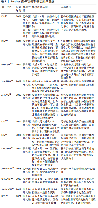
[5] KIM HK, SU PH, QIU YS. Histopathologic changes in growth-plate cartilage following ischemic necrosis of the capital femoral epiphysis. An experimental investigation in immature pigs. J Bone Joint Surg Am. 2001;83(5):688-697.
[6] JOHNSON CP, WANG L, TOTH F, et al. Quantitative susceptibility mapping detects neovascularization of the epiphyseal cartilage after ischemic injury in a piglet model of legg-calve-perthes disease .J Magn Reson Imaging. 2019;50(1):106-113.
[7] ARUWAJOYE OO, MONTE F, KIM A, et al. A comparison of transphyseal neck-head tunneling and multiple epiphyseal drilling on femoral head healing following ischemic osteonecrosis: an experimental investigation in immature pigs. J Pediatr Orthop. 2020;40(4):168-175.
[8] REN Y, DENG Z, GOKANI V, et al. Anti-interleukin-6 therapy decreases hip synovitis and bone resorption and increases bone formation following ischemic osteonecrosis of the femoral head. J Bone Miner Res. 2021;36(2):357-368.
[9] KIM HK, ARUWAJOYE O, DU J, et al. Local administration of bone morphogenetic protein-2 and bisphosphonate during non-weight-bearing treatment of ischemic osteonecrosis of the femoral head: an experimental investigation in immature pigs. J Bone Joint Surg Am. 2014;96(18):1515-1524.
[10] SHAPIRO F, CONNOLLY S, ZURAKOWSKI D, et al. Acetabular changes associated with avascularnecrosis of the femoral head in a piglet model. Bone Joint Res. 2014;3(4):130-138.
[11] ZHANG P, LIANG Y, KIM H, et al. Evaluation of a pig femoral head osteonecrosis model. J Orthop Surg Res. 2010;5:15.
[12] UPASANI VV, JEFFORDS ME, FARNSWORTH CL, et al. Ischemic femoral head osteonecrosis in a piglet model causes three dimensional decrease in acetabular coverage. J Orthop Res. 2018;36(4):1173-1177.
[13] SHAPIRO F, CONNOLLY S, ZURAKOWSKI D, et al. Femoral head deformation and repair following induction of ischemic necrosis: a histologic and magnetic resonance imaging study in the piglet. J Bone Joint Surg Am. 2009;91(12):2903-2914.
[14] JOHNSON CP, WANG L, TOTH F, et al. Quantitative MRI helps to detect hip ischemia: preclinical model of legg-calve-perthes disease. Radiology. 2018;289(2):386-395.
[15] KIM HK, SU PH. Development of flattening and apparent fragmentation following ischemic necrosis of the capital femoral epiphysis in a piglet model. J Bone Joint Surg Am. 2002;84(8):1329-1334.
[16] PRINGLE D, KOOB TJ, KIM HK. Indentation properties of growing femoral head following ischemic necrosis. J Orthop Res. 2004;22(1):122-130.
[17] KIM HK, ARUWAJOYE O, STETLER J, et al. Effects of non-weight-bearing on the immature femoral head following ischemic osteonecrosis: an experimental investigation in immature pigs. J Bone Joint Surg Am. 2012;94(24):2228-2237.
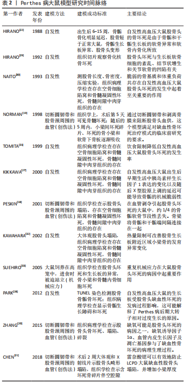
[18] PESKIN B, SHUPAK A, MISSELEVICH I, et al. Transphyseal osseous bridges in experimental osteonecrosis of the femoral head of the rat. Histologic study of the bony bridges connecting the epiphyseal with the metaphyseal bony trabeculae through gaps in the physeal cartilage. J Pediatr Orthop B. 2001;10(3):214-218.
[19] NORMAN D, REIS D, ZINMAN C, et al. Vascular deprivation-induced necrosis of the femoral head of the rat. An experimental model of avascular osteonecrosis in the skeletally immature individual or Legg-Perthes disease. Int J Exp Pathol. 1998;79(3):173-181.
[20] ZHANG W, YUAN Z, PEI X, et al. In vivo and in vitro characteristic of HIF-1alpha and relative genes in ischemic femoral head necrosis. Int J Clin Exp Pathol. 2015;8(6):7210-7216.
[21] CHEN YP, TAN A, HO WP, et al. Effectiveness of strontium ranelate in the treatment of rat model of legg-calve-perthes disease. Indian J Orthop. 2018;52(4):380-386.
[22] HIRANO T, IWASAKI K, YAMANE Y. Osteonecrosis of the femoral head of growing, spontaneously hypertensive rats. Acta Orthop Scand. 1988; 59(5):530-535.
[23] SUEHIRO M, HIRANO T, MIHARA K, et al. Etiologic factors in femoral head osteonecrosis in growing rats. J Orthop Sci. 2000;5(1):52-56.
[24] ZENMYO M, KOMIYA S, KAWABATA R, et al. Morphological and biochemical evidence for apoptosis in the terminal hypertrophic chondrocytes of the growth plate. J Pathol. 1996;180(4):430-433.
[25] KIM H, WENGER D. “Functional retroversion” of the femoral head in Legg-Calvé-Perthes disease and epiphyseal dysplasia: analysis of head-neck deformity and its effect on limb position using three-dimensional computed tomography. J Pediatr Orthop. 1997;17(2):240-246.
[26] KONG SY, KIM HW, PARK HW, et al. Effects of multiple drilling on the ischemic capital femoral epiphysis of immature piglets. Yonsei Med J. 2011;52(5):809-817.
[27] KIKKAWA M, IMAI S, HUKUDA S. Altered postnatal expression of insulin-like growth factor-I (IGF-I) and type X collagen preceding the Perthes’ disease-like lesion of a rat model. J Bone Miner Res. 2000;15(1):111-119.
[28] PARK H, KONG SY, KIM HW. Altered cellular kinetics in the growth plate of the femoral head of spontaneously hypertensive rats. Yonsei Med J. 2012;53(3):625-633.
[29] HIRANO T, IWASAKI K, ODA J, et al. Osteonecrosis of the femoral head in spontaneously hypertensive rats. Relation to ossific nuclei during growth. Acta Orthop Scand. 1992;63(1):37-40.
[30] NAITO S, ITO M, SEKINE I, et al. Femoral head necrosis and osteopenia in stroke-prone spontaneously hypertensive rats (SHRSPs). Bone. 1993; 14(5):745-753.
[31] TOMITA M, SHIMOKAWA I, MAEDA H, et al. Dietary restriction reduces the prevalence of osteonecrosis of the caput femoris in spontaneously hypertensive rats. Calcif Tissue Int. 1999;64(3):259-262.
[32] KAWAHARA T, SHIMOKAWA I, TOMITA M, et al. Effects of caloric restriction on development of the proximal growth plate and metaphysis of the caput femoris in spontaneously hypertensive rats: microscopic and computer-assisted image analyses. Microsc Res Tech. 2002;59(4):306-312.
[33] SUEHIRO M, HIRANO T, SHINDO H. Osteonecrosis induced by standing in growing Wistar Kyoto rats. J Orthop Sci. 2005;10(5):501-507.
[34] BERTHAUME MA, PERRY DC, DOBSON CA, et al. Skeletal immaturity, rostral sparing, and disparate hip morphologies as biomechanical causes for Legg-Calve-Perthes’ disease. Clin Anat. 2016;29(6):759-772.
[35] YAMAGUCHI R, KAMIYA N, KUROYANAGI G, et al. Development of a murine model of ischemic osteonecrosis to study the effects of aging on bone repair. J Orthop Res. 2021. doi: 10.1002/jor.25006.
[36] KAMIYA N, KUROYANAGI G, ARUWAJOYE O, et al. IL6 receptor blockade preserves articular cartilage and increases bone volume following ischemic osteonecrosis in immature mice. Osteoarthritis Cartilage. 2019;27(2):326-335.
[37] KAMIYA N, KIM H. Elevation of proinflammatory cytokine hmgb1 in the synovial fluid of patients with legg-calvé-perthes disease and correlation with IL-6. JBMR Plus. 2020;5(2):e10429.
[38] KAMIYA N, YAMAGUCHI R, ARUWAJOYE O, et al. Development of a mouse model of ischemic osteonecrosis. Clin Orthop Relat Res. 2015; 473(4):1486-1498.
[39] COLE H, YUASA M, HAWLEY G, et al. Differential development of the distal and proximal femoral epiphysis and physis in mice. Bone. 2013; 52(1):337-346.
[40] KUROYANAGI G, ADAPALA NS, YAMAGUCHI R, et al. Interleukin-6 deletion stimulates revascularization and new bone formation following ischemic osteonecrosis in a murine model. Bone. 2018;116:221-231.
[41] PHIPPS M, HUANG Y, YAMAGUCHI R, et al. In vivo monitoring of activated macrophages and neutrophils in response to ischemic osteonecrosis in a mouse model. J Orthop Res. 2016;34(2):307-313.
[42] KAMIYA N, SHAFER S, OXENDINE I, et al. Acute BMP2 upregulation following induction of ischemic osteonecrosis in immature femoral head. Bone. 2013;53(1):239-247.
[43] WANG ZL, HE RZ, TU B, et al. Drilling combined with adipose-derived stem cells and bone morphogenetic protein-2 to treat femoral head epiphyseal necrosis in juvenile rabbits. Curr Med Sci. 2018;38(2):277-288.
[44] ROWE SM, CHUNG JY, MOON ES, et al. The effects of subluxation of the femoral head with avascular necrosis in growing rabbits. J Pediatr Orthop. 2004;24(6):645-650.
[45] 牒军,张开放,王坤正.Perthes病早期骨核素显像的变化[J].中华临床医药杂志(北京),2001,2(11):41-43.
[46] 张开放,李政,姚永锋,等.股骨头骨骺缺血性坏死模型兔构建及组织病理学变化和p53与bcl-2的表达[J].中国组织工程研究与临床康复,2011,15(2):219-223.
[47] 刘浩.Notch信号通路受体及配体在幼兔激素性股骨头缺血坏死模型中的表达及意义[D].遵义:遵义医科大学,2020.
[48] 卓金伟.Wnt/β-catenin信号通路在幼兔激素性股骨头缺血坏死模型中的表达及意义[D].遵义:遵义医学院,2015.
[49] 王健.死亡受体通路相关蛋白在激素性股骨头缺血坏死动物模型中的表达[D].遵义:遵义医学院,2014.
[50] ZHANG JF, YANG CJ, WU T, et al. A two-degree-of-freedom hip exoskeleton device for an immature animal model of exercise-induced Legg-Calve-Perthes disease. Proc Inst Mech Eng H. 2009;223(8):1059-168.
[51] HULTH A, NORBERG I, OLSSON S. Coxa plana in the dog. J Bone Joint Surg Am. 1962;44-A:918-930.
[52] AGUADO E, GOYENVALLE E. Legg Calvé Perthes disease in the dog. Morphologie. 2021;105(349):143-147.
[53] ALPASLAN AM, AKSOY MC, YAZICI M. Interruption of the blood supply of femoral head:an experimental study on the pathogenesis of Legg-Calve-Perthes Disease. Arch Orthop Trauma Surg. 2007;127(6):485-491.
[54] 汪逸,张媛媛,黄绍义,等.骨髓间充质干细胞及其分泌因子对治疗犬股骨头缺血性坏死的效果观察[J].中国兽医杂志,2019,55(8):7.
[55] 官建中,周建生,肖玉周,等.建立幼犬股骨头坏死模型的实验研究[J].蚌埠医学院学报,2006,31(5):444-446.
[56] 闫宏伟,王坤正,时志斌,等.Legg-Perthes 病动物模型设计与评价[J].中国矫形外科杂志,2002,10(8):795-796.
[57] 孟海涛,李林,王翠苹,等.Ⅱ型胶原基因型变化与Legg-perthes病发病的相关性研究[J]实用骨科杂志,2015,21(2):134-135.
[58] ZAHIR A, FREEMAN AR. Cartilage changes following a single episode of infarction of the capital femoral epiphysis in the dog. J Bone Joint Surg Am. 1972;54(1):125-136.
[59] NEWTON B, CRAWFORD CJ, POWERS DL, et al. The immature goat as an animal model for Legg-Calve-Perthes disease. J Invest Surg. 1994; 7(5):417-430.
[60] CRAWFORD CJ, LABERGE M, ALLEN BL JR, et al. Growth profiles and articular cartilage characterization in a goat model of Legg-Calve-Perthes disease. J Invest Surg. 1995;8(6):391-408.
[61] MARTINEZ-ALVAREZ S, EPELDEGUI-TORRE T, MANSO-DIAZ G, et al. Experimental induction of Perthes disease in lambs. Rev Esp Cir Ortop Traumatol. 2014;58(2):68-77.
|