[1] 王璐,张立宁,何家乐,等.不同时长的静态进展性牵伸对大鼠创伤性膝关节挛缩的治疗效果及其机制研究[J].四川大学学报(医学版),2020,51(2):182-192.
[2] CHEN AF, LEE YS, ADAM J, et al. Arthrofibrosis and large joint scarring. Connect Tissue Res. 2019;60(1):21-28.
[3] 关骅.中国骨科康复学[M].北京:人民军医出版社,2011.
[4] YERCAN HS, SUGUN TS, BUSSIERE C, et al. Stiffness after total knee arthroplasty: prevalence, management and outcomes. Knee. 2006; 13(2):111-117.
[5] CANADIAN ORTHOPAEDIC TRAUMA SOCIETY. Open reduction and internal fixation compared with circular fixator application for bicondylar tibial plateau fractures. Results of a multicenter, prospective, randomized clinical trial. J Bone Joint Surg Am. 2006;88(12):2613-2623.
[6] 夏冬瑞,颜继英,张学斌,等.胫骨平台骨折术后膝关节僵硬的发生率及危险因素分析[J].中国骨与损伤杂志,2019,34(12):1315-1317.
[7] USUBA M, MIYANAGA Y, MIYAKAWA S, et al. Effect of heat in increasing the range of knee motion after the development of a joint contracture: an experiment with an animal model. Arch Phys Med Rehabil. 2006; 87(2):247-253.
[8] 武蒙.体外冲击波在大鼠膝关节僵硬治疗中的应用及其作用机制的研究[D].沈阳:中国医科大学,2020.
[9] TRUDEL G, ZHOU J, UHTHOFF HK, et al. Four weeks of mobility after 8 weeks of immobility fails to restore normal motion: a preliminary study. Clin Orthop Relat Res. 2008;466(5):1239-1244.
[10] KANEGUCHI A, OZAWA J, KAWAMATA S, et al. Development of arthrogenic joint contracture as a result of pathological changes inremobilized rat knees. J Orthop Res. 2017;35(7):1414-1423.
[11] KANEGUCHI A, OZAWA J, MINAMIMOTO K, et al. Morphological and biomechanical adaptations of skeletal muscle in the recovery phase after immobilization in a rat. Clin Biomech (Bristol, Avon). 2020;75: 104992.
[12] 古哈尔·艾思拉洪.综合康复护理干预对关节挛缩动物模型形态学及血流动力影响[D].乌鲁木齐:新疆医科大学,2020.
[13] ZHOU HD, TRUDEL G, GOUDREAU L, et al. Knee joint stiffness following immobilization and remobilization: a study in the rat model. J Biomech. 2020;99:109471.
[14] ZHOU HD, TRUDEL G, LANEUVILLE O. Mechanical adaptation of synoviocytes A and B to immobilization and remobilization: a study in the rat knee flexion model. J Mol Histol. 2020;51(5):605-611.
[15] BARLOW JD, HARTZLER RU, ABDEL MP, et al. Surgical capsular release reduces flexion contracture in a rabbit model of arthrofibrosis. J Orthop Res. 2013;31(10):1529-1532.
[16] FUKUI N, FUKUDA A, KOJIMA K, et al. Suppression of fibrous adhesion by proteoglycan decorin. J Orthop Res. 2001;19(3):456-462.
[17] BARLOW JD, MORREY ME, HARTZLER RU, et al. Effectiveness of rosiglitazone in reducing flexion contracture in a rabbit model of arthrofibrosis with surgical capsular release: a biomechanical, histological, and genetic analysis. Bone Joint Res. 2016;5(1):11-17.
[18] BARANOWSKI A, SCHLEMMER L, FÖRSTER K, et al. A novel rat model of stable posttraumatic joint stiffness of the knee. J Orthop Surg Res. 2018;13(1):185.
[19] 王飞,刘克敏,刘四海.康复治疗对创伤后膝关节僵直活动度及肌肉组织的影响[J].中国矫形外科杂志,2017,25(6):534-538.
[20] 黄哲元,袁志,卫磊.山羊膝关节僵直动物模型的建立[J].创伤外科杂志,2010,12(2):159-162.
[21] MONUMENT MJ, HART DA, BEFUS AD, et al. The mast cell stabilizer ketotifen fumarate lessens contracture severity and myofibroblast hyperplasia: a study of a rabbit model of posttraumatic joint contractures. J Bone Joint Surg Am. 2010;92(6):1468-1477.
[22] NESTERENKO S, MORREY S, ABDEL MP, et al. New rabbit knee model of post-traumatic joint contracture: indirect capsular damage induces a severe contracture. Orthop Res. 2009;27(8):1028-1032.
[23] BARANOWSKI A, SCHLEMMER L, FÖRSTER K, et al. Effects of losartan and atorvastatin on the development of early posttraumatic joint stiffness in a rat model. Drug Des Devel Ther. 2019;13:2603-2618.
[24] HAZLEWOOD D, FENG Y, LU QH, et al. Novel rabbit model of moderate knee contracture induced by direct capsular damage. J Orthop Res. 2018;36(10):2687-2695.
[25] SASABE R, SAKAMOTO J, GOTO K, et al. Effects of joint immobilization on changes in myofibroblasts and collagen in the rat knee contracture model. J Orthop Res. 2017;35(9):1998-2006.
[26] SOTOBAYASHI D, KAWAHATA H, ANADA N, et al. Therapeutic effect of intra-articular injection of ribbon-type decoy oligonucleotides for hypoxia inducible factor-1 on joint contracture in an immobilized knee animal model. J Gene Med. 2016;18(8):180-192.
[27] 张全兵.牵伸联合超短波治疗对兔膝关节挛缩模型中关节功能恢复以及关节囊纤维化影响的初步研究[D].合肥:安徽医科大学, 2018.
[28] KOJIMA S, HOSO M, WATANABE M, et al. Experimental joint immobilization and remobilization in the rats. J Phys Ther Sci. 2014; 26(6):865-871.
[29] YUN Z, QUANBING Z, HUAZHANG Z, et al. Rabbit model of extending knee joint contracture: progression of joint motion restriction and subsequent joint capsule changes after immobilization. J Knee Surg. 2020;33(1):15-21.
[30] 张理平,刘晓颖,林绍彬,等.创伤性膝关节僵直动物模型的研究[J].中国中医骨伤科杂,2008,16(5):25-27.
[31] 王锋,徐勇,夏云建.外伤性膝关节僵硬模型兔中药熏洗治疗后内侧副韧带组织中糖胺聚糖的表达[J].中国组织工程研究与临床康复,2010,14(46):8631-8634.
[32] TRUDEL G, UHTHOFF HK. Contractures secondary to immobility: is the restriction articular or muscular? An experimental longitudinal study in the rat knee. Arch Phys Med Rehabil. 2000;81(1):6-13.
[33] HILDEBRAND KA, SUTHERLAND C, MEI Z. Rabbit knee model of post-traumatic joint contractures: the long-term natural history of motion loss and myofibroblasts. J Orthop Res. 2004;22(8):313-320.
[34] HAGIWARA Y, SAIJO Y, CHIMOTO E, et al. Increased elasticity of capsule after immobilization in a rat knee experimental model assessed by scanning acoustic microscopy. Ups J Med Sci. 2006;111(3):303-313.
[35] ABDEL MP, MORREY ME, BARLOW JD, et al. Myofibroblast cells are preferentially expressed early in a rabbit model of joint contracture. J Orthop Res. 2012;30(5):713-719.
[36] KANEGUCHI A, OZAWA J, YAMAOKA K. Anti-inflammatory drug dexamethasone treatment during the remobilization period improves range of motion in a rat knee model of joint contracture. Inflammation. 2018;41(4):1409-1423.
[37] WONG K, TRUDEL G, LANEUVILLE O. Intra-articular collagenase injection increases range of motion in a rat knee flexion contracture model. Drug Des Devel Ther. 2017;12:15-24.
[38] HAGIWARA Y, ANDO A, CHIMOTO E, et al. Expression of collagen types i and ii on articular cartilage in a rat knee contracture model. Connect Tissue Res. 2010;51(1):22-30.
[39] NAGAI M, AOYAMA T, ITO A, et al. Contributions of biarticular myogenic components to the limitation of the range of motion after immobilization of rat knee joint. BMC Musculoskelet Disord. 2014;15: 224.
[40] KANEGUCHI A, OZAWA J, MINAMIMOTO K, et al. Active exercise on immobilization-induced contractured rat knees develops arthrogenic joint contracture with pathological changes. J Appl Physiol. 2018; 124(2):291-301.
[41] ANDO A, SUDA H, HAGIWARA Y, et al. Reversibility of immobilization-induced articular cartilage degeneration after remobilization in rat knee joints. Tohoku J Exp Med. 2011;224(2):77-85.
[42] NAGAI M, ITO A, TAJINO J, et al. Remobilization causes site-specific cyst formation in immobilization-induced knee cartilage degeneration in an immobilized rat model. J Anat. 2016;228(6):929-939.
[43] HAGIWARA Y, ANDO A, CHIMOTO E, et al. Changes of articular cartilage after immobilization in a rat knee contracture model. J Orthop Res. 2009;27(2):236-242.
[44] TRUDEL G, LANEUVILLE O, COLETTA E, et al. Quantitative and temporal differential recovery of articular and muscular limitations of knee joint contractures: results in a rat model. J Appl Physiol. 2014;117(7):730-737.
[45] TRUDEL G, UHTHOFF HK, GOUDREAU L, et al. Quantitative analysis of the reversibility of knee flexion contractures with time: an experimental study using the rat model. BMC Musculoskelet Disord. 2014;15:338.
[46] 王锋.超短波对兔伸直型膝关节挛缩模型中关节功能障碍和废用性肌萎缩治疗作用的初步研究[D].合肥:安徽医科大学,2019.
[47] HAGIWARA Y, ANDO A, ONODA Y, et al. Expression patterns of collagen types i and iii in the capsule of a rat knee contracture model. J Orthop Res. 2010;28(3):315-321.
[48] OAKLEY SP, LASSERE MN, PORTEK I, et al. Biomechanical histologic and macroscopic assessment of articular cartilage in a sheep model of osteoarthritis. Osteoarthritis Cartilage. 2004;12(8):667-679.
[49] OZAWA J, KANEGUCHI A, TANAKA R, et al. Cyclooxygenase-2 inhibitor celecoxib attenuates joint contracture following immobilization in rat knees. BMC Musculoskelet Disord. 2016;17(1):446.
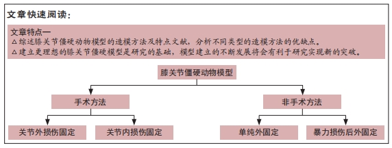
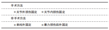
|