中国组织工程研究 ›› 2022, Vol. 26 ›› Issue (24): 3858-3864.doi: 10.12307/2022.566

• 骨髓干细胞 bone marrow stem cells • 上一篇    下一篇

转录因子配对盒基因6对骨髓间充干细胞向角膜缘干细胞样细胞分化的影响

吴  霜,邹星星,高  杰,胡  蓉,苏  敏   

  1. 贵州医科大学基础医学院组织学与胚胎学教研室,贵州省贵阳市   550025
  • 收稿日期:2020-11-14 接受日期:2021-02-27 出版日期:2022-08-28 发布日期:2022-01-22
  • 通讯作者: 苏敏,博士,教授,贵州医科大学基础医学院组织学与胚胎学教研室,贵州省贵阳市 550025
  • 作者简介:吴霜,女,1989年生,贵州省贵阳市人,汉族,2022年贵州医科大学毕业,硕士,主要从事干细胞基础与临床研究。
  • 基金资助:
    贵州省科技支撑项目(黔科合支撑[2020]4Y230号),项目负责人:苏敏;贵州科技厅人才平台计划(黔科合平台人才[2020]4103号),项目负责人:苏敏;贵州省科学技术项目(黔科合基础[2020]1Y408),项目负责人:高杰;贵州省科技厅学术新苗培养项目(黔科合平台人才[2018]5779-21),项目负责人:高杰;贵州省教育厅青年科技人才成长项目(黔教合KY字[2018]201),项目负责人:高杰

Effect of paired box gene 6 protein on the differentiation of bone marrow mesenchymal stem cells into keratolimbal stem cell-like cells

Wu Shuang, Zou Xingxing, Gao Jie, Hu Rong, Su Min   

  1. Department of Histology and Embryology, College of Basic Medical Sciences, Guizhou Medical University, Guiyang 550025, Guizhou Province, China
  • Received:2020-11-14 Accepted:2021-02-27 Online:2022-08-28 Published:2022-01-22
  • Contact: Su Min, MD, Professor, Department of Histology and Embryology, College of Basic Medical Sciences, Guizhou Medical University, Guiyang 550025, Guizhou Province, China
  • About author:Wu Shuang, Master, Department of Histology and Embryology, College of Basic Medical Sciences, Guizhou Medical University, Guiyang 550025, Guizhou Province, China
  • Supported by:
    Guizhou Science and Technology Support Project, No. [2020]4Y230 (to SM); Talent Platform Program of Guizhou Science and Technology Department, No. [2020]4103 (to SM); Guizhou Science and Technology Project, No. [2020]1Y408) (to GJ); Academic New Seedling Training Project of Guizhou Provincial Science and Technology Department, No. [2018]5779-21) (to GJ); Youth Science and Technology Talent Growth Project of Guizhou Education Department, No. KY[2018]201 (to GJ)

摘要:

文题释义:
配对盒基因6:在脊椎动物眼睛发育过程中是必不可少的,在人类怀孕的第5周,配对盒基因6存在于视网膜的神经层和色素层,也在来自表面外胚层的前段结构中高度表达,包括晶状体囊泡和角膜上皮。出生后,配对盒基因6仅限于视网膜神经节、无长突细胞和水平细胞、晶状体、角膜、结膜、虹膜和睫状体。
角膜缘干细胞:角膜缘为角膜和结膜、巩膜交界部分,具有不断增殖分化和向心性移动的能力,能够修复替代衰老死亡的角膜上皮细胞,在角膜上皮的更新和角膜疾病的治疗中起着不可替代的作用,其属于单能干细胞,在角膜损伤修复中发挥重要作用。

背景:角膜病移植手术面临着角膜供者严重缺乏、术后发生免疫排异反应等诸多问题。在大力倡导角膜捐献的同时,急需找到一种新的替代性的治疗手段。
目的:通过优化骨髓间充质干细胞培养条件,探讨转录因子配对盒基因6对骨髓间充质干细胞向角膜缘干细胞样细胞分化的影响。
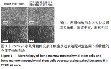
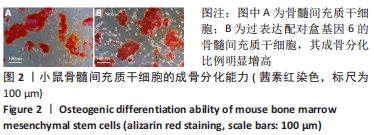
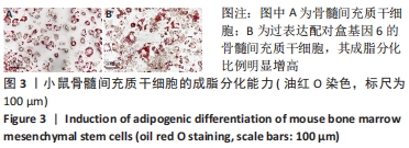
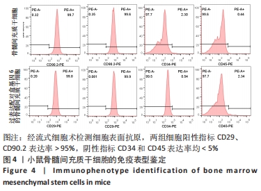
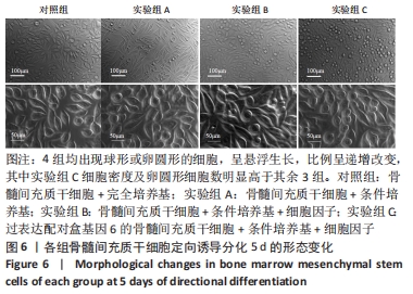
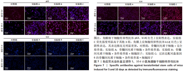
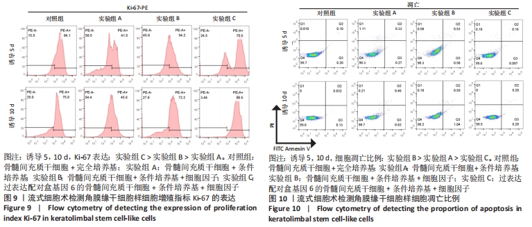
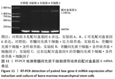
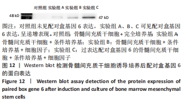
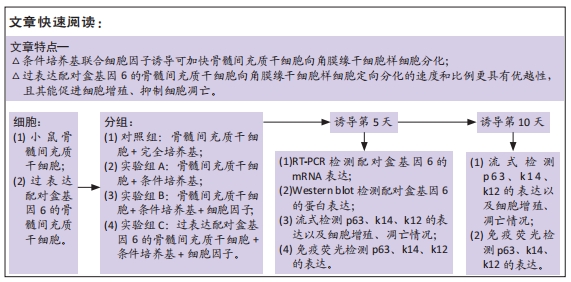
方法:根据细胞形态、成骨成脂分化能力、细胞表面抗原表达鉴定骨髓间充质干细胞及过表达配对盒基因6的骨髓间充质干细胞。按照培养细胞和培养基不同进行分组:对照组(骨髓间充质干细胞+完全培养基)、实验组A(骨髓间充质干细胞+条件培养基)、实验组B(骨髓间充质干细胞+条件培养基+细胞因子)、实验组C(过表达配对盒基因6的骨髓间充质干细胞+条件培养基+细胞因子)。诱导5,10 d时采用免疫荧光、流式细胞术检测p63、k14和k12的表达,流式细胞术检测细胞增殖指标Ki-67的表达和细胞凋亡率,Western blot法和RT-PCR法检测配对盒基因6的蛋白和mRNA表达。
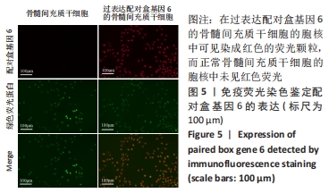
结果与结论:①诱导培养前,两组细胞均具有骨髓间充质干细胞的特性。②诱导培养后,均出现球型或卵圆形的细胞,呈悬浮生长。4组细胞的角膜缘干细胞样细胞特异性分子p63、k14表达阳性,实验组荧光表达量高于对照组(P < 0.05),而角膜上皮分子k12表达阴性。随着培养条件的优化,实验组C的细胞增殖更明显,凋亡细胞更少。诱导5 d时,实验各组配对盒基因6的蛋白及mRNA表达量呈递增变化。③结果表明,条件培养基联合细胞因子诱导可加快骨髓间充质干细胞向角膜缘干细胞样细胞分化。过表达配对盒基因6的骨髓间充质干细胞向角膜缘干细胞样细胞定向分化的速度和比例更具有优越性,且其能促进细胞增殖、抑制细胞凋亡。
缩略语:骨髓间充质干细胞:bone marrow mesenchymal stem cells,BM-MSCs;角膜缘干细胞:limbal stem cells,LSCs

https://orcid.org/0000-0001-9867-9952 (吴霜) 

中国组织工程研究杂志出版内容重点:干细胞;骨髓干细胞;造血干细胞;脂肪干细胞;肿瘤干细胞;胚胎干细胞;脐带脐血干细胞;干细胞诱导;干细胞分化;组织工程

关键词: 干细胞, 骨髓间充干细胞, 角膜缘干细胞, 配对盒基因6, 细胞因子, 条件培养基

Abstract: BACKGROUND: Keratopathy transplantation is faced with many problems, such as serious shortage of corneal donors, postoperative immune rejection and so on. While vigorously advocating corneal donation, there is an urgent need to find a new alternative treatment.  
OBJECTIVE: The effect of paired box gene 6 on the differentiation of bone marrow mesenchymal stem cells into keratolimbal stem cell-like cells was investigated by optimizing the culture conditions of bone marrow mesenchymal stem cells.
METHODS:  Bone marrow mesenchymal stem cells and bone marrow mesenchymal stem cells overexpressing paired box gene 6 were identified according to cell morphology, osteogenic adipogenic differentiation ability, and cell surface antigen expression. According to different cultured cells and culture medium, they were divided into control group (bone marrow mesenchymal stem cells + complete medium), and experimental group A (bone marrow mesenchymal stem cells + conditioned medium), experimental group B (bone marrow mesenchymal stem cells + conditioned medium + cytokine), and experimental group C (bone marrow mesenchymal stem cells overexpressing paired box gene 6 + conditioned medium + cytokine). The expression levels of p63, k14 and k12 were detected by immunofluorescence and flow cytometry at 5 and 10 days after induction. The expression of Ki-67, an indicator of cell proliferation, and the rate of apoptosis, were measured by flow cytometry. Western blot assay and RT-PCR were used to determine the protein and mRNA expression of paired box gene 6.  
RESULTS AND CONCLUSION: (1) Before induction and culture, the two groups of cells had the characteristics of bone marrow mesenchymal stem cells. (2) After conditioned culture, spherical or oval cells appeared in the four groups after induction, which showed suspension growth. The expression of keratolimbal stem cell-like cells molecules p63 and k14 was positive in the four groups. The fluorescence expression in the experimental group was significantly higher than that in the control group (P < 0.05), but the expression of corneal epithelial molecule k12 was negative. With the optimization of the culture conditions, the cell proliferation of experimental group C was more obvious, and there were fewer apoptotic cells. After 5 days of induction, the expression of paired box gene 6 protein and mRNA in the experimental groups increased progressively at 5 days after induction. (3) Results suggested that conditioned medium combined with cytokine induction can accelerate the differentiation of bone marrow mesenchymal stem cells into keratolimbal stem cell-like cells. Bone marrow mesenchymal stem cells overexpressing paired box gene 6 are more superior in the speed and proportion of directional differentiation into keratolimbal stem cell-like cells, and it can promote cell proliferation and inhibit cell apoptosis.

Key words: stem cells, bone marrow mesenchymal stem cells, keratolimbal stem cell-like cells, paired box gene 6, cytokines, conditioned medium

中图分类号: