中国组织工程研究 ›› 2021, Vol. 25 ›› Issue (33): 5249-5253.doi: 10.12307/2021.309

• 数字化骨科 digital orthopedics •    下一篇

3D打印技术辅助经颈前路的新型椎弓根固定系统治疗颈椎病合并骨质疏松的三维有限元分析

杨九杰1,2,王  涛1,李  治1,杨立枫1,田  野3,毕  征4,曾雅玲2   

  1. 1沈阳医学院附属中心医院骨科,辽宁省沈阳市   110034;2澳门科技大学,中国澳门特别行政区;3北京协和医院骨科,北京市   100000;4北京市医用内植物工程技术研究中心,北京市   100082
  • 收稿日期:2021-03-16 修回日期:2021-04-06 接受日期:2021-04-14 出版日期:2021-11-28 发布日期:2021-08-02
  • 通讯作者: 李治,医学博士,主任医师,沈阳医学院附属中心医院骨科,辽宁省沈阳市 110034
  • 作者简介:杨九杰,1992年生,汉族,澳门科技大学在读博士。
  • 基金资助:
    辽宁省科技厅重点研发计划项目基金(2017225066),项目负责人:李治

Cervical spondylosis with osteoporosis treated by the new pedicle fixation system through the anterior cervical approach with three-dimensional printing technology: three-dimensional finite element analysis

Yang Jiujie1, 2, Wang Tao1, Li Zhi1, Yang Lifeng1, Tian Ye3, Bi Zheng4, Zeng Yaling2   

  1. 1Department of Orthopedics, Affiliated Central Hospital of Shenyang Medical College, Shenyang 110034, Liaoning Province, China; 2Macao University of science and technology, Macao Special Administration Region, China; 3Department of Orthopedics, Peking Union Medical College Hospital, Beijing 100000, China; 4Beijing Medical Implant Engineering Technology Research Center, Beijing 100082, China
  • Received:2021-03-16 Revised:2021-04-06 Accepted:2021-04-14 Online:2021-11-28 Published:2021-08-02
  • Contact: Li Zhi, MD, Chief physician, Department of Orthopedics, Affiliated Central Hospital of Shenyang Medical College, Shenyang 110034, Liaoning Province, China
  • About author:Yang Jiujie, Doctoral candidate, Department of Orthopedics, Affiliated Central Hospital of Shenyang Medical College, Shenyang 110034, Liaoning Province, China; Macao University of science and technology, Macao Special Administration Region, China
  • Supported by:
    Key Research and Development Project Fund of Liaoning Provincial Department of Science and Technology, No. 2017225066 (to LZ)

摘要:


文题释义:

颈椎病合并骨质疏松:骨质疏松可导致人的颈椎变形,然后造成椎管狭窄,是促进颈椎发生退行性变化进而发展为颈椎病的一种潜在的危险因素。
骨质疏松症:是一种以低骨量和骨组织微结构破坏为特征,导致骨质脆性增加和易于骨折的全身性骨代谢性疾病。
背景:骨质疏松患者颈椎内固定治疗的稳定性差,骨折愈合过程缓慢,同一部位及其他部位发生再骨折的风险明显增大,因此需要设计出一种新的经前路内固定的方式,以期减少并发症及手术翻修率。
目的:采用三维有限元分析和3D打印技术,设计新型治疗颈椎病合并骨质疏松经前路椎弓根入路的固定系统,有利于提高手术后患者颈椎的稳定性。
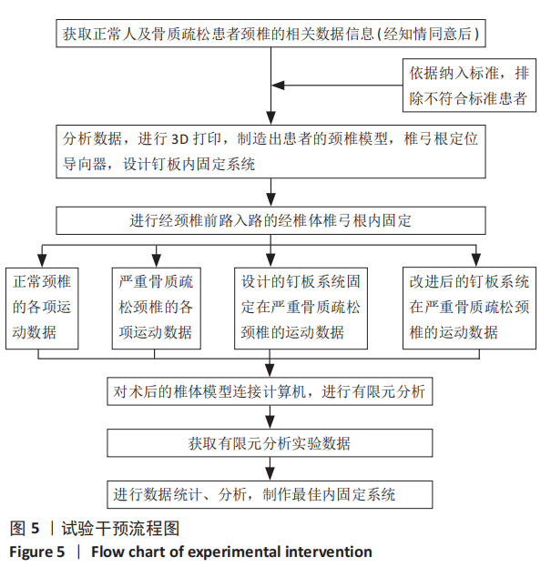
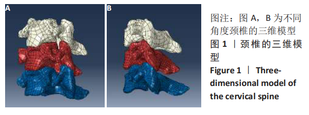
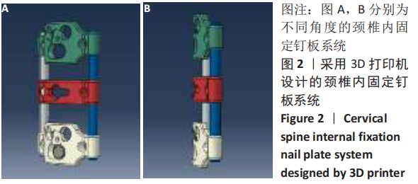
方法:收集2017年12月至2019年12月在沈阳医学院附属中心医院骨科体检的15例颈椎健康者和15例骨质疏松患者的三维CT数据资料,通过3D打印技术构建30例受试者颈椎的三维模型,依据椎弓根定位导向器,设计出新型钉板内固定系统,并对3D模型进行体外条件下定位器引导的颈椎前路入路的椎体椎弓根固定。随后将制备好的术后模型连接计算机,进行有限元分析及生物力学分析,依据分析结果设计出最佳钉板内固定系统。

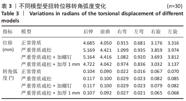
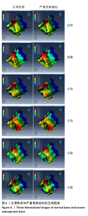
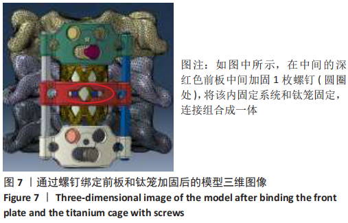
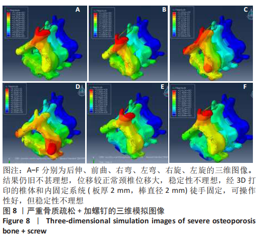
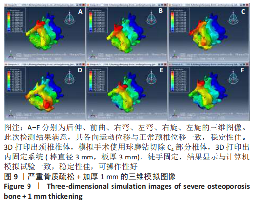
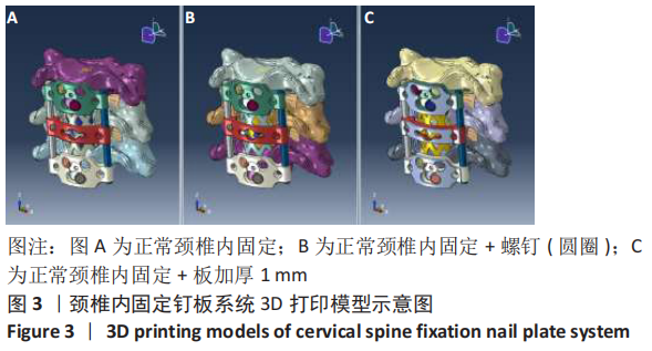
结果与结论:①利用3D打印技术成功构建出骨质疏松患者的颈椎模型,以及经椎体前方椎弓根固定定位导向器和经椎体前方椎弓根固定钉板系统;②有限元生物力学分析结果显示,严重骨质疏松患者骨质比正常骨质受扭转位移和转角明显增高,加螺钉对承受前后曲和左右弯扭转变形改善不明显,加螺钉对承受轴向扭转载荷变形有明显改善;③结果证实,试验借助3D打印技术设计出一种用于颈椎前路入路的椎体椎弓根内固定钉板系统,证实3D打印技术可以成功辅助颈椎经前路椎弓根固定来治疗颈椎病合并骨质疏松。

https://orcid.org/0000-0002-9487-2920 (李治) 

中国组织工程研究杂志出版内容重点:人工关节;骨植入物;脊柱;骨折;内固定;数字化骨科;组织工程

关键词: ">骨, 颈椎, 3D打印技术, 骨质疏松, 椎弓根, 固定, 有限元分析

Abstract: BACKGROUND: In osteoporosis patients, the stability of cervical spine after internal fixation is poor; the fracture healing process is slow; and the risk of re-fracture at the same site and other sites is significantly increased. Therefore, it is necessary to design a new method of anterior internal fixation to reduce complications and surgical revision rate.  
OBJECTIVE: To design a new fixation system for the treatment of cervical spondylosis with osteoporosis through the anterior pedicle approach using three-dimensional (3D) finite element analysis and 3D printing technology, which is beneficial to improve the stability of the patient’s cervical spine after surgery.
METHODS:  3D CT data of 15 healthy cervical spine patients and 15 osteoporosis patients who were examined in the Department of Orthopedics, Affiliated Central Hospital of Shenyang Medical College from December 2017 to December 2019 were collected. The cervical spine models of 30 patients were constructed by 3D printing technology. A new nail plate internal fixation system was designed based on the pedicle positioning guide. 3D models were subjected to the vertebral pedicle fixation through the anterior approach guided by the localizer under external conditions. The prepared postoperative models were connected to the computer for finite element analysis and biomechanical analysis, and the best nail plate internal fixation system was designed based on the analysis results.  
RESULTS AND CONCLUSION: (1) 3D printing technology was used to successfully construct the cervical spine models of osteoporosis patients, as well as the anterior vertebral pedicle fixation and positioning guide and the anterior vertebral pedicle fixation nail plate system. (2) Finite element biomechanics analysis results showed that the bone mass of patients with severe osteoporosis was significantly higher than that of normal bone by torsional displacement and rotation angle. The addition of screws did not significantly improve the torsional deformation of the anterioposterior bending and the left and right bending. The addition of screws had a significant improvement in the deformation of the axial torsional load. (3) The results indicated that the trial designed a vertebral pedicle internal fixation nail plate system for the anterior cervical approach using 3D printing technology, confirming that 3D printing technology can successfully assist the cervical spine through anterior pedicle fixation for cervical spondylosis combined with osteoporosis.

Key words: bone, cervical spine, 3D printing technology, osteoporosis, pedicle, fixation, finite element analysis

中图分类号: